. 2012 Apr 6;287(15):12395-404.
doi: 10.1074/jbc.M111.306530. Epub 2012 Jan 10.

Endotoxin priming of neutrophils requires endocytosis and NADPH oxidase-dependent endosomal reactive oxygen species

Affiliations

Endotoxin priming of neutrophils requires endocytosis and NADPH oxidase-dependent endosomal reactive oxygen species

Fred S Lamb et al. J Biol Chem. .

Abstract

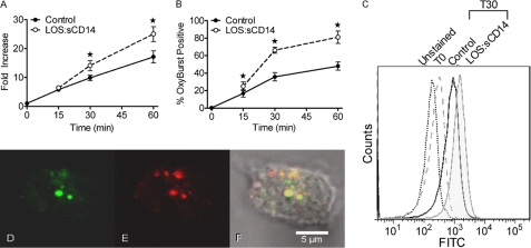
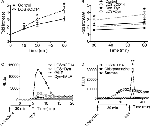
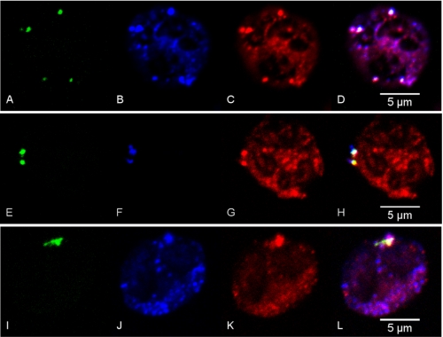
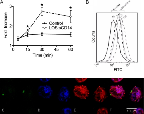
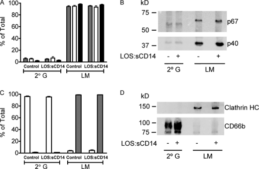
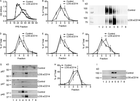
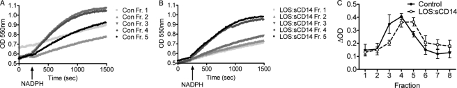
NADPH oxidase 2 (Nox2)-generated reactive oxygen species (ROS) are critical for neutrophil (polymorphonuclear leukocyte (PMN)) microbicidal function. Nox2 also plays a role in intracellular signaling, but the site of oxidase assembly is unknown. It has been proposed to occur on secondary granules. We previously demonstrated that intracellular NADPH oxidase-derived ROS production is required for endotoxin priming. We hypothesized that endotoxin drives Nox2 assembly on endosomes. Endotoxin induced ROS generation within an endosomal compartment as quantified by flow cytometry (dihydrorhodamine 123 and Oxyburst Green). Inhibition of endocytosis by the dynamin-II inhibitor Dynasore blocked endocytosis of dextran, intracellular generation of ROS, and priming of PMN by endotoxin. Confocal microscopy demonstrated a ROS-containing endosomal compartment that co-labeled with gp91(phox), p40(phox), p67(phox), and Rab5, but not with the secondary granule marker CD66b. To further characterize this compartment, PMNs were fractionated by nitrogen cavitation and differential centrifugation, followed by free flow electrophoresis. Specific subfractions made superoxide in the presence of NADPH by cell-free assay (cytochrome c). Subfraction content of membrane and cytosolic subunits of Nox2 correlated with ROS production. Following priming, there was a shift in the light membrane subfractions where ROS production was highest. CD66b was not mobilized from the secondary granule compartment. These data demonstrate a novel, nonphagosomal intracellular site for Nox2 assembly. This compartment is endocytic in origin and is required for PMN priming by endotoxin.


Figures

FIGURE 1.
FIGURE 1.
Endotoxin priming elicits intracellular ROS production. A, intracellular ROS were measured by flow cytometry using the cell permeable DHR 123 fluorescent ROS-sensitive probe. LOS:sCD14 10 ng/ml, elicited a time-dependent increase in intracellular ROS as compared to control. Control cells at time 0, immediately following addition of the probe, are set to a value of 1, and data are presented as fold-increase with respect to this value, * = p < 0.05, n = 10. B, using Oxyburst to measure intracellular ROS in endocytic compartments, LOS:sCD14-primed PMNs displayed a significantly greater percentage of Oxyburst positive cells than did control PMN, * = p < 0.05, n = 6. C, representative histogram from compiled data in B. D–F, confocal microscopy demonstrated Oxyburst positive vesicular compartments in a subset of cells. Representative confocal image of PMN after 30 min of priming with LOS:sCD14 demonstrating Oxyburst positive, ROS-containing vesicles (D), colocalizing with TR-dextran containing vesicles (E), and merged image (F).
FIGURE 2.
FIGURE 2.
Endotoxin priming elicits endocytosis, and endocytosis is required for priming of the respiratory burst. A, flow cytometry analysis of endocytosis of TR-dextran in control versus LOS:sCD14-treated PMN demonstrates a significant increase in endocytosis in response to priming. Fold increase presented with respect to control cells at time 0, * = p < 0.05, n = 5. B, effect of the dynamin-II inhibitor Dynasore on endocytosis of TR dextran in control versus LOS:sCD14 PMNs. Dynasore (Dyn, 300 μm) significantly inhibits constitutive endocytosis in unstimulated PMNs. Dynasore also blocked LOS:sCD14-mediated enhancement of TR-dextran endocytosis, * = p < 0.05, n = 6. C, PMN were primed with LOS:sCD14 in the absence or presence of Dynasore for 30 min before washing twice and then stimulating with fMLF. Primed NADPH oxidase activity, as measured by LUC-CL, in response to fMLF was markedly inhibited in the cells primed in the presence of Dynasore, representative tracing, n = 8. D, both chlorpromazine (40 μm) and sucrose (225 mm) significantly inhibited LOS:sCD14-primed NADPH oxidase activity, as measured by LUC-CL, in response to fMLF. ** = p < 0.05 sucrose compared to LOS:sCD14 without inhibitor, * = p < 0.05 for chlorpromazine compared to no inhibitor, n = 8.
FIGURE 3.
FIGURE 3.
Confocal microscopy analysis of endotoxin primed PMN displays ROS-containing vesicles that colocalize with NADPH oxidase components and early endosomal markers. PMNs were incubated for 30 min with LOS:sCD14 (10 ng/ml) in the presence of the ROS-sensitive probe, Oxyburst, and Alexa Fluor 647 dextran. Oxyburst positive, ROS-containing vesicles are seen in green (A, E, I) and colocalize with gp91phox in blue (B, J), Alexa Fluor 647 dextran in blue (F), p40phox (C), p67phox (G), and rab5 (K), all in red. D, H, L, representative merged images.
FIGURE 4.
FIGURE 4.
Endotoxin priming elicits low-level mobilization of secondary granules to the cell surface, but ROS-containing endosomes do not express secondary granule markers. A, cell surface levels of CD66b are significantly enhanced by priming with LOS:sCD14, 10 ng/ml, as compared to control conditions, * = p < 0.05, n = 9. B, representative flow cytometry histogram of CD66b. C–F, confocal microscopy of endotoxin primed PMNs display Oxyburst positive, ROS-containing vesicles (C). Robust staining for the secondary granule markers (D) CD66b (blue), and (E) lactoferrin (red) is present in all cells, but there is no colocalization of ROS-positive vesicles with either CD66b or lactoferrin (F). Representative merged image is from 4 independent experiments.
FIGURE 5.
FIGURE 5.
NADPH oxidase cytosolic subunits are associated with the light membrane (LM) fraction, but not the secondary granule (2° G) fraction, isolated from both control and endotoxin-primed PMN. A–B, control and LOS:sCD14-primed (10 ng/ml) PMN were subjected to cavitation and Percoll density gradient centrifugation with isolation of each of the granule fractions. LM and 2° G subfractions were immunoblotted for p40phox (grey-filled bars), p47phox (open bars), and p67phox (black bars). Greater than 90% of the Nox2 cytosolic subunit proteins, which were membrane-associated, were found associated with the LM fraction. There was no increase in cytosolic subunit abundance associated with the LM subfraction following endotoxin priming. Data were compiled from n = 5–6 individual experiments. B, representative immunoblot displaying p67phox and p40phox. C, immunoblotting for the 2° G marker CD66b (open bars) displayed minimal mobilization from the 2° G fraction to the LM fraction after priming with LOS:sCD14. Immunoblotting for clathrin heavy chain (grey-filled bars) demonstrated significant association of clathrin with the LM fraction but none associated with the 2° G compartment, n = 4. D, representative immunoblot displaying clathrin HC and CD66b.
FIGURE 6.
FIGURE 6.
Endotoxin priming of PMN elicits an alteration in the properties of the light membrane subfractions. A, alkaline phosphatase activity assay performed on the FFE subfractions from control and endotoxin-primed, 10 ng/ml LOS:sCD14. PMN demonstrates a shift in the fractions displaying alkaline phosphatase activity post-Triton X-100 permeabilization, representative activity assay, from n = 5 individual experiments. FFE subfractions were pooled into groups of 8 for further biochemical analysis. B–C, gp91phox protein was detected primarily in pooled subfractions 3, 4, and 5 from control PMNs, but shifted to fractions 4, 5, and 6 following endotoxin priming. B, compiled data from n = 5 separate FFE runs. C, representative immunoblot for gp91phox. D–F, cytosolic subunits of the oxidase were also associated with light membrane pooled subfractions isolated by FFE. p40phox (D) and p67phox (F) displayed similar shifts by immunoblotting as seen with gp91phox, whereas p47phox (E) had only a minor shift following priming, n = 5. G, representative immunoblot for control versus LOS:sCD14 primed PMNs for cytosolic subunits of Nox2. H–I, there was no shift in the clathrin heavy chain protein content of FFE subfractions following endotoxin priming, n = 5.
FIGURE 7.
FIGURE 7.
Cell-free NAPDH oxidase assay for measurement of superoxide production using the reduction of ferricytochrome c displays a shift in the superoxide generating fractions following endotoxin priming. A, in control PMN, pooled subfractions 3 and 4 generated the greatest amount of superoxide following addition of NADPH, whereas peak superoxide generating activity shifted to subfractions 4 and 5 following endotoxin priming (B). C, compiled data for superoxide generation by subfractions from n = 3 FFE experiments comparing control versus endotoxin-primed PMN displays a full fraction shift in peak superoxide generating activity.

References

    1. Bass D. A., Olbrantz P., Szejda P., Seeds M. C., McCall C. E. (1986) Subpopulations of neutrophils with increased oxidative product formation in blood of patients with infection. J. Immunol. 136, 860–866 - PubMed
    1. Chollet-Martin S., Montravers P., Gibert C., Elbim C., Desmonts J. M., Fagon J. Y., Gougerot-Pocidalo M. A. (1992) Subpopulation of hyperresponsive polymorphonuclear neutrophils in patients with adult respiratory distress syndrome. Role of cytokine production. Am. Rev. Respir. Dis. 146, 990–996 - PubMed
    1. Ogura H., Tanaka H., Koh T., Hashiguchi N., Kuwagata Y., Hosotsubo H., Shimazu T., Sugimoto H. (1999) Priming, second-hit priming, and apoptosis in leukocytes from trauma patients. J. Trauma 46, 774–783 - PubMed
    1. Condliffe A. M., Kitchen E., Chilvers E. R. (1998) Neutrophil priming. Pathophysiological consequences and underlying mechanisms. Clin. Sci. 94, 461–471 - PubMed
    1. Swain S. D., Rohn T. T., Quinn M. T. (2002) Neutrophil priming in host defense. Role of oxidants as priming agents. Antioxid. Redox Signal. 4, 69–83 - PubMed

Publication types

MeSH terms