

In vivo MRI characterization of progressive cardiac dysfunction in the mdx mouse model of muscular dystrophy
- PMID: 22235247
- PMCID: PMC3250389
- DOI: 10.1371/journal.pone.0028569
In vivo MRI characterization of progressive cardiac dysfunction in the mdx mouse model of muscular dystrophy
Abstract
Aims: The mdx mouse has proven to be useful in understanding the cardiomyopathy that frequently occurs in muscular dystrophy patients. Here we employed a comprehensive array of clinically relevant in vivo MRI techniques to identify early markers of cardiac dysfunction and follow disease progression in the hearts of mdx mice.
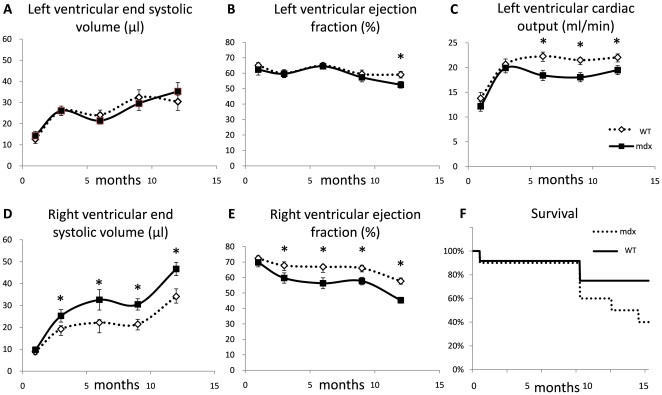
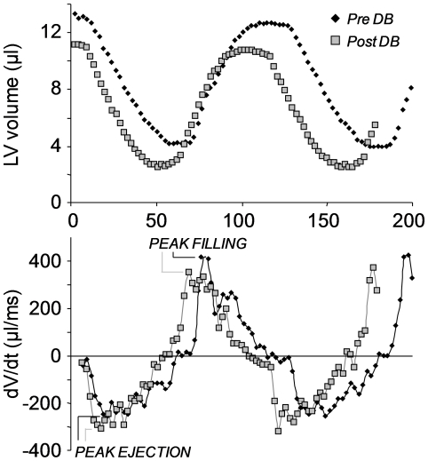
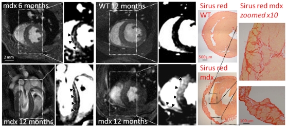
Methods and results: Serial measurements of cardiac morphology and function were made in the same group of mdx mice and controls (housed in a non-SPF facility) using MRI at 1, 3, 6, 9 and 12 months after birth. Left ventricular (LV) and right ventricular (RV) systolic and diastolic function, response to dobutamine stress and myocardial fibrosis were assessed. RV dysfunction preceded LV dysfunction, with RV end systolic volumes increased and RV ejection fractions reduced at 3 months of age. LV ejection fractions were reduced at 12 months, compared with controls. An abnormal response to dobutamine stress was identified in the RV of mdx mice as early as 1 month. Late-gadolinium-enhanced MRI identified increased levels of myocardial fibrosis in 6, 9 and 12-month-old mdx mice, the extent of fibrosis correlating with the degree of cardiac remodeling and hypertrophy.
Conclusions: MRI could identify cardiac abnormalities in the RV of mdx mice as young as 1 month, and detected myocardial fibrosis at 6 months. We believe these to be the earliest MRI measurements of cardiac function reported for any mice, and the first use of late-gadolinium-enhancement in a mouse model of congenital cardiomyopathy. These techniques offer a sensitive and clinically relevant in vivo method for assessment of cardiomyopathy caused by muscular dystrophy and other diseases.
Conflict of interest statement
Figures


References
-
- Finsterer J, Stollberger C. The heart in human dystrophinopathies. Cardiology. 2003;99:1–19. - PubMed
-
- AMERICAN-ACADEMY-OF-PEDIATRICS. Cardiovascular health supervision for individuals affected by Duchenne or Becker muscular dystrophy. Pediatrics. 2005;116:1569–1573. - PubMed
-
- Lapidos KA, Kakkar R, McNally EM. The dystrophin glycoprotein complex: signaling strength and integrity for the sarcolemma. Circ Res. 2004;94:1023–1031. - PubMed
-
- Fayssoil A, Nardi O, Orlikowski D, Annane D. Cardiomyopathy in Duchenne muscular dystrophy: pathogenesis and therapeutics. Heart Fail Rev. 2010;15:103–107. - PubMed
Publication types
MeSH terms
Substances
Grants and funding
LinkOut - more resources
Full Text Sources
Medical
Molecular Biology Databases
