. 2012;7(1):e29239.
doi: 10.1371/journal.pone.0029239. Epub 2012 Jan 3.

Receptor heteromerization expands the repertoire of cannabinoid signaling in rodent neurons

Affiliations

Receptor heteromerization expands the repertoire of cannabinoid signaling in rodent neurons

Raphael Rozenfeld et al. PLoS One. 2012.

Abstract

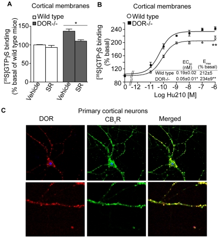
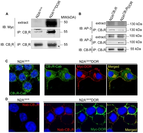
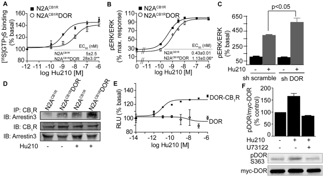
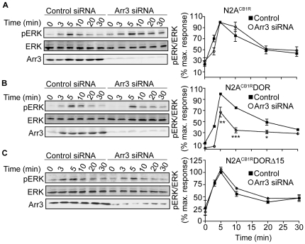
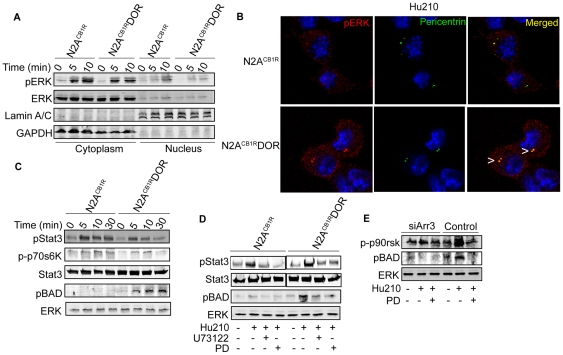
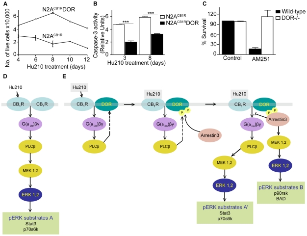
A fundamental question in G protein coupled receptor biology is how a single ligand acting at a specific receptor is able to induce a range of signaling that results in a variety of physiological responses. We focused on Type 1 cannabinoid receptor (CB₁R) as a model GPCR involved in a variety of processes spanning from analgesia and euphoria to neuronal development, survival and differentiation. We examined receptor dimerization as a possible mechanism underlying expanded signaling responses by a single ligand and focused on interactions between CB₁R and delta opioid receptor (DOR). Using co-immunoprecipitation assays as well as analysis of changes in receptor subcellular localization upon co-expression, we show that CB₁R and DOR form receptor heteromers. We find that heteromerization affects receptor signaling since the potency of the CB₁R ligand to stimulate G-protein activity is increased in the absence of DOR, suggesting that the decrease in CB₁R activity in the presence of DOR could, at least in part, be due to heteromerization. We also find that the decrease in activity is associated with enhanced PLC-dependent recruitment of arrestin3 to the CB₁R-DOR complex, suggesting that interaction with DOR enhances arrestin-mediated CB₁R desensitization. Additionally, presence of DOR facilitates signaling via a new CB₁R-mediated anti-apoptotic pathway leading to enhanced neuronal survival. Taken together, these results support a role for CB₁R-DOR heteromerization in diversification of endocannabinoid signaling and highlight the importance of heteromer-directed signal trafficking in enhancing the repertoire of GPCR signaling.


Conflict of interest statement

Competing Interests: The authors have declared that no competing interests exist.

Figures

Figure 1
Figure 1. Increased CB1R activity in the absence of DOR.
A, Basal [35S]GTPγS binding was measured in cortical membranes from wild-type and DOR −/− mice. Membranes from cortices were prepared as described –, treated with vehicle or 1 µM SR141716 (SR) for 1 hour and subjected to [35S]GTPγS binding as described in Methods. Basal [35S]GTPγS binding/10 µg protein in vehicle treated membranes is taken as 100%. Data represent Mean ± SEM (n = 3 individual animals in triplicate). Statistically significant differences between vehicle and SR141716 treatment are indicated *p<0.05, (t test). B, [35S]GTPγS binding assay in cortical membranes from wild-type and DOR −/− mice. Membranes were treated with increasing concentrations of the CB1R agonist Hu210 (0–1 µM) and [35S]GTPγS binding was measured as described in Methods. EC50 and Emax values were calculated using GraphPad Prism software. Data represent Mean ± SEM (n = 3 individual animals in triplicate). *p<0.05; **p<0.01 for DOR−/− vs wild-type (t test). C, Localization of endogenous CB1R and DOR in mouse primary cortical cells, 14DIV. Cells fixed with 4%PFA in PBS and permeablized with 0.1% Triton, were immunostained with the goat polyclonal anti-CB1R(N-terminal) antibody (1∶500; green) and rat polyclonal anti-DOR antibody (1∶500; red) and visualized using Alexa 488-conjugated anti-goat (1∶1000) and Alexa 594-conjugated anti-rat (1∶1000) secondary antibodies using confocal microscopy as described in Methods. Representative figure from 3 independent experiments shown.
Figure 2
Figure 2. Association between CB1R and DOR alters CB1R localization.
A, Lysates (100–200 µg) from N2ACB1R and N2ACB1RDOR cells were subjected to immunoprecipitation with 1 µg of anti-CB1R (C-terminal) antibody, the immunoprecipitates were resolved on 10% SDS-PAGE and probed for the presence of myc-DOR using mouse monoclonal anti-myc antibody (1∶1000) and for CB1R using rabbit polyclonal anti-CB1R (C-terminal) antibody (1∶500) as described in Methods. IRDye 680 anti-rabbit and IRDye 800 anti-mouse were used as secondary antibodies (1∶10,000). Representative of 3 independent experiments shown. B, CB1R-DOR complexes exhibit greater interaction with AP-2 than AP-3. Lysates (100–200 µg) from N2ACB1R and N2ACB1RDOR cells were subjected to immunoprecipitation using 1 µg of an anti-CB1R (C-terminal) antibody as described in Methods. The immunoprecipitates were resolved on 10% SDS-PAGE and probed for the presence of AP-3 (1∶1000), AP-2 (1∶1000) and CB1R (C-term) (1∶500) using specific antibodies as described in Methods. IRDye 680 anti-rabbit and IRDye 800 anti-mouse were used as secondary antibodies (1∶10,000). Representative of 3 independent experiments shown. C, Localization of endogenous CB1R in N2ACB1R and of CB1R and DOR in N2ACB1RDOR cells. Cells fixed with 4%PFA in PBS and permeablized with 0.1% Triton, were stained with the rabbit polyclonal anti-CB1R (C-terminal) antibody (1∶500; green) and the mouse monoclonal anti-myc antibody (1∶1000; red) and visualized using Alexa 488-coupled anti-rabbit or Alexa 594-coupled anti-mouse secondary antibodies (1∶1000) using confocal microscopy as described in Methods. Representative of 3 independent experiments shown. D, Cell surface staining of endogenous CB1R and stably expressed DOR in N2ACB1RDOR cells. N2ACB1R and N2ACB1RDOR cells were stained with a goat polyclonal anti-CB1R (N-terminal) antibody (1∶500) and mouse monoclonal anti-myc antibodies (1∶1000) prior to fixation of the cells to label cell surface receptors, as described . After fixation, cells were visualized with Alexa 594-coupled anti-goat and Alexa 488-coupled anti-mouse secondary antibodies (1∶1,000) using confocal microscopy as described in Methods. Representative of 3 independent experiments shown.
Figure 3
Figure 3. PLC-dependent arrestin3 association with CB1R-DOR complex.
A, [35S]GTPγS binding assay in membranes from N2ACB1R and N2ACB1RDOR cells. Membranes (10 µg) were treated with indicated concentrations of the CB1R agonist Hu210. [35S]GTPγS binding was measured as described in Methods. EC50 and Emax values were calculated using GraphPad Prism software. Data represent Mean ± SEM (n = 3 independent experiments in triplicate). B, Dose-response of Hu210-mediated ERK phosphorylation in N2ACB1R and N2ACB1RDOR cells. Starved N2ACB1R and N2ACB1RDOR cells seeded in 24 well-plates were treated with indicated concentrations of Hu210 for 5 minutes. Cell lysates (30 µg protein) were analyzed by Western blotting and probed for the levels of pERK (1∶1000) and ERK (1∶1000) as described in Methods. IRDye 680 anti-rabbit and IRDye 800 anti-mouse were used as secondary antibodies (1∶10,000). EC50 and Emax values were calculated using GraphPad Prism software. Data represent Mean ± SEM (n = 3 independent experiments). *p<0.05 for N2ACB1RDOR vs N2ACB1R (t test). C, Effect of DOR down-regulation on ERK phosphorylation. F11 cells transduced with the DOR shRNA expressing lentivirus were starved for 4–6 h and treated with Hu210 (100 nM) for 5 min. Cell lysates (30 µg protein) were analyzed by Western blotting and probed for the levels of pERK (1∶1000) and ERK (1∶1000) as described in Methods. IRDye 680 anti-rabbit and IRDye 800 anti-mouse were used as secondary antibodies (1∶10,000). Data from 3 independent experiments is shown. *p<0.05 (t test). D, Examination of arrestin3 interaction with CB1R after Hu210 treatment. N2ACB1R and N2ACB1RDOR, starved for 4 hours were stimulated with 100 nM Hu210 for 5 minutes and cell lysates prepared as described in Methods. Lysates (30 µg protein) were subjected to either Western blotting using rabbit anti-CB1R (C-terminal 1∶500) and mouse anti-arrestin 3 antibodies (1∶500) or to immunoprecipitation using 1 µg of agarose-coupled anti-CB1R (C-terminal) antibody. Immunoprecipitates were probed for arrestin3 levels by Western blot using the mouse anti-arrestin 3 antibody. IRDye 680 anti-rabbit and IRDye 800 anti-mouse were used as secondary antibodies (1∶10,000). Representative of 3 independent experiments shown. E, Effect of Hu210 on arrestin recruitment. U2OS cells co-expressing ProLink/Enzyme Donor (PK)-tagged DOR and the Enzyme Activator (EA)-tagged arrestin3 fusion protein without or with CB1R were treated with indicated concentrations of Hu-210. Arrestin3 recruitment was determined using the PathHunter Detection Kit as described in Methods. Data represent Mean ± SEM (n = 4). F, Effect of PLC inhibitor (U73122) on DOR phosphorylation at serine 363 after Hu210 treatment. N2ACB1RDOR cells were starved for 4–6 hours, and incubated with vehicle (DMSO) or U73122 (1 µM) for 30 minutes, then stimulated with 100 nM Hu210 for 5 minutes. Cell lysates (30 µg protein) were subjected to Western blotting using rabbit polyclonal phosphoDOR Ser 363 (1∶1000), mouse monoclonal anti-myc (1∶1000) antibodies and IR Dye 680 anti-rabbit and IR Dye 800 anti-mouse secondary antibodies (1∶10,000) as described in Methods. Data represent Mean ± SEM (n = 3).
Figure 4
Figure 4. Engagement of arrestin3-dependent signaling in N2ACB1RDOR.
Time course of Hu210-mediated ERK phosphorylation in A, N2ACB1R; B, N2ACB1RDOR; and C, N2ACB1RDORΔ15 cells transfected with control or arrestin3-targeting siRNA. N2ACB1R alone or stably expressing either DOR or DORΔ15, transfected with a control or arrestin3-targeting siRNA, were starved for 4 hours, then stimulated with 100 nM Hu210 for the indicated times. Cell lysates (30 µg protein) were subjected to Western blotting for the levels of ERK (1∶1000), pERK (1∶1000), and arrestin3 (1∶500) as described in Methods. IRDye 680 anti-rabbit and IRDye 800 anti-mouse were used as secondary antibodies (1∶10,000). Data represent Mean ± SEM (n = 3); *p<0.05; **p<0.01; ***p<0.001, for control vs Arr3 siRNA (t test).
Figure 5
Figure 5. Role of arrestin3 and ERK substrates in cannabinoid signaling by the CB1R-DOR heteromer.
A–B, Effect of CB1R-DOR heteromerization on subcellular localization of pERK. A, N2ACB1R and N2ACB1RDOR cells were treated with Hu210 (100 nM; 0, 5 or 10 min), cytoplasmic and nuclear extracts were prepared as described in Methods and analyzed (30 µg protein) by Western blotting with pERK (1∶1000), ERK (1∶1000), lamin A/C (1∶2000), and GAPDH (1∶2000) antibodies. IRDye 680 anti-rabbit and IRDye 800 anti-mouse were used as secondary antibodies (1∶10,000). B, N2ACB1R and N2ACB1RDOR cells treated with Hu210 (100 nM; 5 min) were immunostained with pERK (1∶1000, red) and pericentrin (1∶1000, green) antibodies and visualized using Alexa 488-conjugated anti-rabbit (1∶1000) and Alexa 594-conjugated anti-mouse (1∶1000) secondary antibodies using confocal microscopy as described in Methods. Representative of 3 independent experiments shown. C, Time course of phosphorylation of pERK substrates. Lysates (30 µg protein) from N2ACB1R and N2ACB1RDOR cells treated with Hu210 (100 nM; 0, 5, 10 or 30 min) were analyzed by Western blotting with STAT3 (1∶1000), phosphoSTAT3 (1∶1000), phospho-p70S6K (1∶1000), phospho-p90rsk (1∶1000) and phosphoBAD (1∶1000) antibodies. IRDye 680 anti-rabbit and IRDye 800 anti-mouse were used as secondary antibodies (1∶10,000). ERK (1∶1000) is used as a loading control. Representative of 3 independent experiments shown. D, Involvement of PLC and MEK in the phosphorylation of STAT3 and BAD. Lysates (30 µg protein) from N2ACB1R and N2ACB1RDOR cells treated with 100 nM Hu210 for 5 min in the absence or presence of U73122 (1 µM) or PD98059 (PD, 10 µM) were analyzed by Western blotting with STAT3 (1∶1000), phosphoSTAT3 (1∶1000), and phosphoBAD (1∶1000) antibodies. IRDye 680 anti-rabbit and IRDye 800 anti-mouse were used as secondary antibodies (1∶10,000). ERK (1∶1000) is used as a loading control. Representative of 3 independent experiments shown. E, Involvement of arrestin3 in BAD phosphorylation. Arrestin3 was down-regulated in N2ACB1RDOR cells by transfection with a siRNA. These cells were stimulated with Hu210 (100 nM) for 5 min, in the absence or presence of PD (10 µM). Lysates (30 µg protein) were analyzed by Western blotting with phospho-p90rsk (1∶1000) and phosphoBAD (1∶1000) antibodies. IRDye 680 anti-rabbit and IRDye 800 anti-mouse were used as secondary antibodies (1∶10,000). ERK (1∶1000) is used as a loading control. Representative of 3 independent experiments shown.
Figure 6
Figure 6. CB1R-DOR heteromerization promotes cell survival.
A, Hu210-treated N2ACB1RDOR cells exhibit increased survival compared to N2ACB1R cells. N2ACB1R or N2ACB1RDOR cells were treated with 1 µM Hu210 for the indicated days and survival measured by trypan blue exclusion as described in Methods. Data represent Mean ± SEM (n = 4 in triplicate). B, Hu210-treated N2ACB1RDOR cells exhibit lower apoptosis as compared to N2ACB1R cells. Apoptosis of N2ACB1R or N2ACB1RDOR treated for 3 or 8 days with 1 µM Hu210 was measured using caspase-3 activity assay as described in Methods. Data represent Mean ± SEM (n = 4 in triplicate); ***p<0.001 N2ACB1RDOR vs N2ACB1R (t test). C, CB1R antagonist treatment decreases neuronal survival of striatal neurons from wild-type but not DOR−/− mice. Primary striatal neurons from wild-type or from DOR−/− mice were prepared as described in Methods. The CB1R antagonist AM251 (10 µM) was added to the growth media at DIV7 and cellular viability assessed at DIV10 as described in Methods. Data represent Mean ± SEM (n = 2–4) D–E, A schematic of the signaling pathways emanating from CB1R in N2ACB1R (D) and N2ACB1RDOR (E) cells. Activation of CB1R in N2ACB1RDOR cells leads to differential activation of signaling molecules and phosphorylation of ERK substrates.

Similar articles

Cited by

References

    1. Berghuis P, Rajnicek AM, Morozov YM, Ross RA, Mulder J, et al. Hardwiring the brain: endocannabinoids shape neuronal connectivity. Science. 2007;316:1212–1216. - PubMed
    1. Harkany T, Keimpema E, Barabas K, Mulder J. Endocannabinoid functions controlling neuronal specification during brain development. Mol Cell Endocrinol. 2008;286:S84–90. - PubMed
    1. Guzman M, Sanchez C, Galve-Roperh I. Cannabinoids and cell fate. Pharmacol Ther. 2002;95:175–184. - PubMed
    1. Bromberg KD, Ma'ayan A, Neves SR, Iyengar R. Design logic of a cannabinoid receptor signaling network that triggers neurite outgrowth. Science. 2008;320:903–909. - PMC - PubMed
    1. Harkany T, Guzman M, Galve-Roperh I, Berghuis P, Devi LA, et al. The emerging functions of endocannabinoid signaling during CNS development. Trends Pharmacol Sci. 2007;28:83–92. - PubMed

Publication types

MeSH terms

LinkOut - more resources