. 2012;7(1):e29343.
doi: 10.1371/journal.pone.0029343. Epub 2012 Jan 3.

CART peptide is a potential endogenous antioxidant and preferentially localized in mitochondria

Affiliations

CART peptide is a potential endogenous antioxidant and preferentially localized in mitochondria

Peizhong Mao et al. PLoS One. 2012.

Abstract

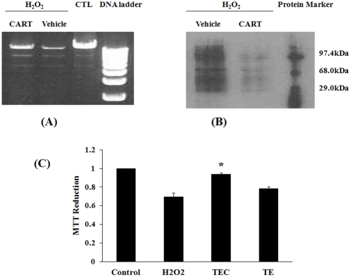
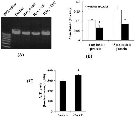
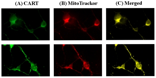
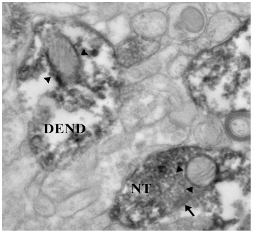
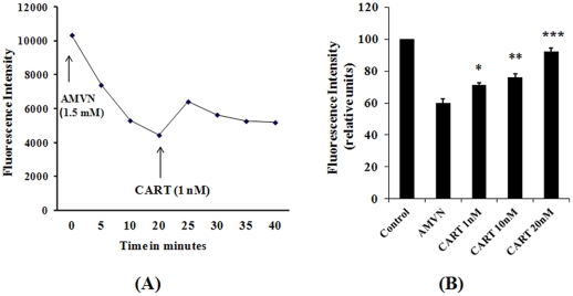
The multifunctional neuropeptide Cocaine and Amphetamine Regulated Transcript (CART) is secreted from hypothalamus, pituitary, adrenal gland and pancreas. It also can be found in circulatory system. This feature suggests a general role for CART in different cells. In the present study, we demonstrate that CART protects mitochondrial DNA (mtDNA), cellular proteins and lipids against the oxidative action of hydrogen peroxide, a widely used oxidant. Using cis-parinaric acid as a sensitive reporting probe for peroxidation in membranes, and a lipid-soluble azo initiator of peroxyl radicals, 2,2'-azobis(2,4-dimethylvaleronitrile) we found that CART is an antioxidant. Furthermore, we found that CART localized to mitochondria in cultured cells and mouse brain neuronal cells. More importantly, pretreatment with CART by systemic injection protects against a mouse oxidative stress model, which mimics the main features of Parkinson's disease. Given the unique molecular structure and biological features of CART, we conclude that CART is an antioxidant peptide (or antioxidant hormone). We further propose that it may have strong therapeutic properties for human diseases in which oxidative stress is strongly involved such as Parkinson's disease.


Conflict of interest statement

Competing Interests: The authors have declared that no competing interests exist.

Figures

Figure 1
Figure 1. CART reduces oxidative damage in HEK293 cells.
(A) CART protects mitochondrial DNA damage induced by H2O2. Total DNA was isolated from HEK293 cells following treatments. Mitochondrial DNAs were amplified by Long PCR. Marker, 1 kb ladder. (B) CART reduces hydrogen peroxide-oxidized proteins. Proteins were damaged by reactive oxygen species (H2O2), vehicle treatment had no effect. However CART fusion protein (TEC 5 µg/ml) decreased the oxidative damage. (C) CART diminishes cell death induced by hydrogen peroxide. Cell viability was monitored by mitochondrial function assayed by MTT reduction: H2O2 (0.1 mM) caused a decline of cellular MTT reduction, and CART treatment, but not vehicle, significantly increased MTT metabolism. *, P<0.05, comparing with H2O2 group, n = 6.
Figure 2
Figure 2. CART antioxidant role in human lymphocytes.
(A) CART protects mitochondrial DNA damage induced by H2O2. MtDNAs were amplified by Long PCR as described in Fig. 1. (B) CART reduces the levels of lipid peroxidation end products. Protein extracts from lymphocytes treated with 4 and 8 µg TATEGFP-CART were compared to TATEGFP following H2O2 treatment. Total MDA and HNE formation was detected using 4 mg of total cell homogenates as starting material. Absorbance was measured at 586 nm and data are mean±SEM of 4 independent experiments. *, P<0.05, comparing to vehicle group. (C) CART preserves cellular ATP production in human lymphocytes. Cultured cells were pretreated with vehicle or CART fusion protein (5μg/ml) and then treated with H2O2 (0.1 mM) to induce cell death. Cell viability assay performed using a sensitive ATP-based detection method is also an indicator of ATP levels. *, P<0.01, comparing to vehicle group, n = 4.
Figure 3
Figure 3. Mitochondrial localization of CART in primary neuronal cells.
Mouse cortex neurons (upper panel) and hippocampal neurons (bottom panel) at 7-day in vitro were incubated with MitoTracker Red. The cells were fixed and incubated with a CART specific antibody and secondary antibody, finally treated with tyramide-conjugated fluorescent dye Alexa 488 (green) and photographed using a fluorescence microscope at 100× magnification. (A) shows CART antibody staining (green), (B) shows mitochondrial staining (red), and (c) is merged image (yellow), demonstrating the co-localization of CART and mitochondria.
Figure 4
Figure 4. Mitochondrial localization of CART in mouse brain.
In the mouse nucleus accumbens determined by electron microscopy. The dark diaminobenzidene reaction product shows that CART is located in a nerve terminal (NT) making a symmetrical synaptic contact (arrow) onto a neuronal cell body. Within the NT is a mitochondrion in which there is CART labeling (arrowheads) around the outer and the inner mitochondrial membranes. Within the photograph is a CART-labeled dendrite (DEND) in which there is a mitochondrion showing CART labeling, particularly along the outer mitochondrion membrane (arrowheads). Magnification: ×40,000.
Figure 5
Figure 5. Radical scavenging role of CART.
(A) Effects of CART on AMVN-induced fluorescence decay in mitochondria isolated from human SHSY cells. AMVN 1.5 mM was added to the reaction medium (0.2 ml) containing mitochondria 50 µg, cis PA 12 µM in 0.1 M KCl and phosphate buffer (50 mM, pH 7.4 at 40°C). CART (1 nM) was added 20 min later. The fluorescence intensity was monitored by using a BIO-TEK spectrofluorometer. (B) Radical scavenging activity of CART in mitochondria isolated from HEK293 cells. The reaction containing 50 µg mitochondria, cisPA 6 µM, CART peptides and AMVM 2 mM in 0.1 M KCI and phosphate buffer was performed at 40°C for 20 min, and the fluorescence intensity was measured after continued incubation at RT for 90 min. *, P<0.05; **, P<0.01; ***, P<0.001, comparing with AMVM group.
Figure 6
Figure 6. Effect of CART on the number of TH-labeled neuronal cells in the SN-PC.
A.) Example of TH-immunolabeled neurons within the SN-PC in all four groups: CTL: control group (vehicle + vehicle); CART (CART + vehicle); MPTP (vehicle + MPTP); MPTP/CART (CART + MPTP). There is a decrease in the number of TH-labeled cells in the MPTP only group compared to all other groups. Administration of CART reversed the MPTP-induced decrease in TH-labeled cells. B.) CART peptide (50 ng/mouse, IP) or vehicle (0.1 ml/10 grams, IP) was injected one day prior to the start of sub-acute administration of either MPTP (7 mg/kg/d) or vehicle for 7 days. On days 1–7, CART or vehicle were injected 30 minutes prior to administration of either MPTP or vehicle. On day 8, mice were perfused with fixative and the SN-PC cut and processed for TH immunolabeling. CART pretreatment blocked the MPTP-induced decrease in the number of TH labeled cells/section of the SN-PC. *, p<0.05 compared to all other groups.
Figure 7
Figure 7. Effect of CART on motor behavior.
(A) Percent free rears. The number of free versus wall-assisted rears was counted over a 5-minute period on day 8 after the last of the MPTP/CART/vehicle injections and the results presented as percent free rears. For the Control (CTL) group, there was nearly a 50/50 split in terms of the number of free versus wall-assisted rears. There was a decrease in the percent free rears for the CART only group, which was equivalent to that seen in the MPTP treated group. CART pretreatment reversed the MPTP-induced decrease in free rears to the levels seen in the control group. *, p<0.05 compared to the CTL and MPTP/CART groups. (B) Number of foot-faults. The number of foot-faults during a 5-minute period was determined on day 8 after the last of the MPTP/CART/vehicle injections. CART pretreatment, then followed by MPTP, resulted in a significant decrease in the number of foot-faults compared to the MPTP only group. *, p<0.05 compared to the MPTP group. (C) Schematic of mechanisms of CART action. Based on our data, CART peptide functions through at least two different and associated pathways, interaction with mitochondrial SDH and directly scavenging oxidants, ultimately preserving cells/neurons.

References

    1. Reichlin S. Neuroendocrine-immune interactions. N Engl J Med. 1993;329(17):1246–1253. - PubMed
    1. Ojeda SR, Lomniczi A, Mastronardi C, Heger S, Roth C, et al. Minireview: The neuroendocrine regulation of puberty: Is the time ripe for a systems biology approach? Endocrinology. 2006;147(3):1166–1174. - PubMed
    1. McEwen BS. Central effects of stress hormones in health and disease: Understanding the protective and damaging effects of stress and stress mediators. Eur J Pharmacol. 2008;583(2–3):174–185. - PMC - PubMed
    1. Snyder SH. Neurotransmitters, receptors, and second messengers galore in 40 years. J Neurosci. 2009;29(41):12717–12721. - PMC - PubMed
    1. Douglass J, McKinzie AA, Couceyro P. PCR differential display identifies a rat brain mRNA that is transcriptionally regulated by cocaine and amphetamine. J Neurosci. 1995;15(3 Pt 2):2471–2481. - PMC - PubMed

Publication types

MeSH terms

Substances