

Keratinocyte-targeted overexpression of the glucocorticoid receptor delays cutaneous wound healing
- PMID: 22235328
- PMCID: PMC3250471
- DOI: 10.1371/journal.pone.0029701
Keratinocyte-targeted overexpression of the glucocorticoid receptor delays cutaneous wound healing
Abstract
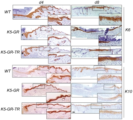
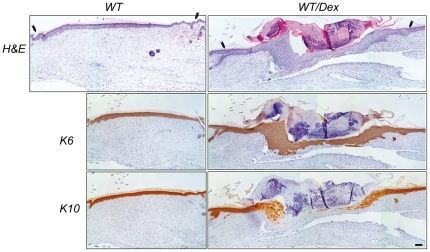
Delayed wound healing is one of the most common secondary adverse effects associated to the therapeutic use of glucocorticoid (GC) analogs, which act through the ligand-dependent transcription factor GC-receptor (GR). GR function is exerted through DNA-binding-dependent and -independent mechanisms, classically referred to as transactivation (TA) and transrepression (TR). Currently both TA and TR are thought to contribute to the therapeutical effects mediated by GR; however their relative contribution to unwanted side effects such as delayed wound healing is unknown. We evaluated skin wound healing in transgenic mice with keratinocyte-restricted expression of either wild type GR or a mutant GR that is TA-defective but efficient in TR (K5-GR and K5-GR-TR mice, respectively). Our data show that at days (d) 4 and 8 following wounding, healing in K5-GR mice was delayed relative to WT, with reduced recruitment of granulocytes and macrophages and diminished TNF-α and IL-1β expression. TGF-β1 and Kgf expression was repressed in K5-GR skin whereas TGF-β3 was up-regulated. The re-epithelialization rate was reduced in K5-GR relative to WT, as was formation of granulation tissue. In contrast, K5-GR-TR mice showed delays in healing at d4 but re-established the skin breach at d8 concomitant with decreased repression of pro-inflammatory cytokines and growth factors relative to K5-GR mice. Keratinocytes from both transgenic mice closed in vitro wounds slower relative to WT, consistent with the in vivo defects in cell migration. Overall, the delay in the early stages of wound healing in both transgenic models is similar to that elicited by systemic treatment with dexamethasone. Wound responses in the transgenic keratinocytes correlated with reduced ERK activity both in vivo and in vitro. We conclude that the TR function of GR is sufficient for negatively regulating early stages of wound closure, while TA by GR is required for delaying later stages of healing.
Conflict of interest statement
Figures


Similar articles
-
Transrepression function of the glucocorticoid receptor regulates eyelid development and keratinocyte proliferation but is not sufficient to prevent skin chronic inflammation.Mol Endocrinol. 2008 Apr;22(4):799-812. doi: 10.1210/me.2007-0284. Epub 2008 Jan 3. Mol Endocrinol. 2008. PMID: 18174358 Free PMC article.
-
The suppressor of cytokine signaling (SOCS)-3 determines keratinocyte proliferative and migratory potential during skin repair.J Invest Dermatol. 2010 Mar;130(3):876-85. doi: 10.1038/jid.2009.344. Epub 2009 Nov 19. J Invest Dermatol. 2010. PMID: 19924142
-
Gypenoside LXXV Promotes Cutaneous Wound Healing In Vivo by Enhancing Connective Tissue Growth Factor Levels Via the Glucocorticoid Receptor Pathway.Molecules. 2019 Apr 23;24(8):1595. doi: 10.3390/molecules24081595. Molecules. 2019. PMID: 31018484 Free PMC article.
-
Role of TGF beta-mediated inflammation in cutaneous wound healing.J Investig Dermatol Symp Proc. 2006 Sep;11(1):112-7. doi: 10.1038/sj.jidsymp.5650004. J Investig Dermatol Symp Proc. 2006. PMID: 17069018 Review.
-
Roles of the Glucocorticoid and Mineralocorticoid Receptors in Skin Pathophysiology.Int J Mol Sci. 2018 Jun 29;19(7):1906. doi: 10.3390/ijms19071906. Int J Mol Sci. 2018. PMID: 29966221 Free PMC article. Review.
Cited by
-
Effects of Superficial Scratching and Engineered Nanomaterials on Skin Gene Profiles and Microbiota in SKH-1 Mice.Int J Mol Sci. 2023 Oct 26;24(21):15629. doi: 10.3390/ijms242115629. Int J Mol Sci. 2023. PMID: 37958613 Free PMC article.
-
Nouveau procédé: les greffes séquentielles de cellules cutanées guérissent-elles les brûlures de troisième degré? étude comparative à propos de 517 patients.Ann Burns Fire Disasters. 2018 Sep 30;31(3):213-222. Ann Burns Fire Disasters. 2018. PMID: 30863256 Free PMC article. French.
-
GRowing an epidermal tumor.J Invest Dermatol. 2013 Dec;133(12):2659-2662. doi: 10.1038/jid.2013.350. J Invest Dermatol. 2013. PMID: 24216781 Free PMC article.
-
Epigenetic Regulation of Epidermal Stem Cell Biomarkers and Their Role in Wound Healing.Int J Mol Sci. 2015 Dec 24;17(1):16. doi: 10.3390/ijms17010016. Int J Mol Sci. 2015. PMID: 26712738 Free PMC article. Review.
-
Flightless-I homolog regulates glucocorticoid receptor-mediated transcription via direct interaction of the leucine-rich repeat domain.Mol Biol Rep. 2017 Apr;44(2):243-250. doi: 10.1007/s11033-017-4106-3. Epub 2017 Apr 28. Mol Biol Rep. 2017. PMID: 28455686
References
-
- Schäcke H, Döcke WD, Asadullah K. Mechanisms involved in the side effects of glucocorticoids. Pharmacol Ther. 2002;96:23–43. - PubMed
-
- Barnes PJ, Adcock IM. Glucocorticoid resistance in inflammatory diseases. Lancet. 2009;373:1905–1917. - PubMed
-
- Revollo JR, Cidlowski JA. Mechanisms Generating Diversity in glucocorticoid Receptor Signaling. Ann NY Acad Sci. 2009;1179:167–178. - PubMed
-
- Stanisić V, Lonard DM, O'Malley BW. Modulation of steroid hormone receptor activity. Prog Brain Res. 2010;181:153–176. - PubMed
Publication types
MeSH terms
Substances
LinkOut - more resources
Full Text Sources
Research Materials
Miscellaneous
