. 2012;7(1):e29813.
doi: 10.1371/journal.pone.0029813. Epub 2012 Jan 3.

Melanopsin-expressing amphioxus photoreceptors transduce light via a phospholipase C signaling cascade

Affiliations

Melanopsin-expressing amphioxus photoreceptors transduce light via a phospholipase C signaling cascade

Juan Manuel Angueyra et al. PLoS One. 2012.

Abstract

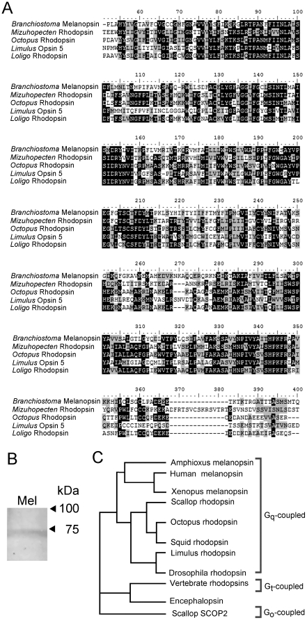
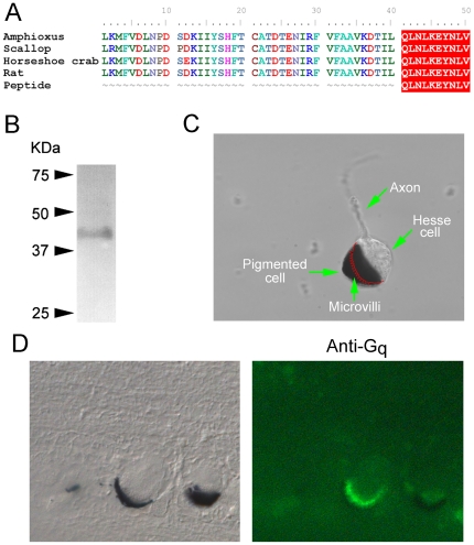
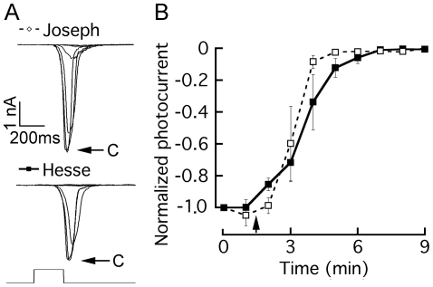
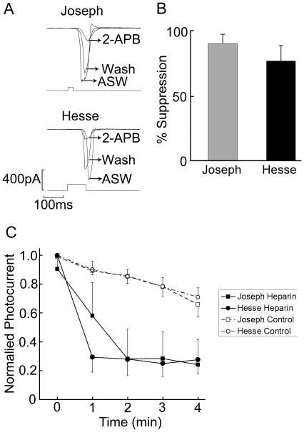
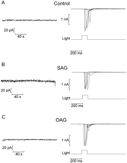
Melanopsin, the receptor molecule that underlies light sensitivity in mammalian 'circadian' receptors, is homologous to invertebrate rhodopsins and has been proposed to operate via a similar signaling pathway. Its downstream effectors, however, remain elusive. Melanopsin also expresses in two distinct light-sensitive cell types in the neural tube of amphioxus. This organism is the most basal extant chordate and can help outline the evolutionary history of different photoreceptor lineages and their transduction mechanisms; moreover, isolated amphioxus photoreceptors offer unique advantages, because they are unambiguously identifiable and amenable to single-cell physiological assays. In the present study whole-cell patch clamp recording, pharmacological manipulations, and immunodetection were utilized to investigate light transduction in amphioxus photoreceptors. A G(q) was identified and selectively localized to the photosensitive microvillar membrane, while the pivotal role of phospholipase C was established pharmacologically. The photocurrent was profoundly depressed by IP₃ receptor antagonists, highlighting the importance of IP₃ receptors in light signaling. By contrast, surrogates of diacylglycerol (DAG), as well as poly-unsaturated fatty acids failed to activate a membrane conductance or to alter the light response. The results strengthen the notion that calcium released from the ER via IP₃-sensitive channels may fulfill a key role in conveying--directly or indirectly--the melanopsin-initiated light signal to the photoconductance; moreover, they challenge the dogma that microvillar photoreceptors and phoshoinositide-based light transduction are a prerogative of invertebrate eyes.


Conflict of interest statement

Competing Interests: The authors have declared that no competing interests exist.

Figures

Figure 1
Figure 1. Amphioxus melanopsin groups with Gq-coupled rhodopsins.
(A) The translated sequence of Branchiostoma was subjected to a BLAST search, and subsequently aligned with the highest-ranking hits among non-melanopsin photopigments. All belong to microvillar photoreceptors from eyes of invertebrates. The Clustal W alignment shows the core region of the proteins (omitting the carboxy-terminus, and part of the amino-terminus, which are generally divergent); black shading indicates identity, whereas grey shading indicates conservative aminoacid substitutions. Accession numbers: amphioxus (Branchiostoma) Q4R1I4; scallop (Mizuhopecten), O15973.1, octopus (Octopus). P09241.1; horseshoe crab (Limulus), ACO05013.1; and squid (Loligo) P24603.1. (B) Western blot with anti-melanopsin antibodies. A single band of ≈71 kDa was detected, closely approaching the size expected from the predicted aminoacid sequence. This confirms that the melanopsin protein expresses in the neural tube of B. floridae. (C) Phylogenetic tree illustrating the grouping of the main classes of photopigments found in the animal kingdom. Amphioxus melanopsin is closely related to the Gq-coupled rhodopsin of mollusks and arthropods.
Figure 2
Figure 2. Gq expresses in the microvillar membrane of Hesse cells.
(A) The sequence of the immunogenic peptide used to raise anti-Gq antibodies was aligned with the predicted C-terminal region of Gq of amphioxus, and those of other organisms. (B) Western blot of neural tube using anti Gq, showing the detection of a single band of ≈42 kDa. (C) Morphological characteristics of the Hesse cell: the accessory pigmented cell engulfs the microvilli-covered region of the clear sensory cell; the position of the villous membrane within the occluded region is drawn in red. (D) Left: Nomarski micrograph of a 10 µm section of fixed neural tube containing two Hesse cells. Right: fluorescence image of the same section incubated with anti-Gq antibodies and Alexa Fluo 488-conjugated secondary antibodies. The immunostaining is confined to the region of the microvilli.
Figure 3
Figure 3. Pharmacological interference with PLC suppresses the photocurrent.
The PLC antagonist U-73122 (10 µM) was applied to voltage-clamped photoreceptors stimulated every minute with a flash of light of fixed intensity. (A) Superimposed current traces measured in a Joseph cell and in a Hesse cell; after photocurrent stabilization, two control responses were measured (‘c’ and arrows), and then the drug was introduced by local superfusion, inducing a progressive decline in the peak amplitude of the light-evoked current in both cell types. (B) Time course of the effect of the PLC inhibitor, averaged for 4 Hesse cells (solid lines/filled symbols) and 3 Joseph cells (dashed lines/open symbols); error bars indicate standard error; the starting time of drug application is marked by the arrow.
Figure 4
Figure 4. Inhibition of the light response by antagonists of the IP3 receptor.
(A) Effect of 2-APB applied extracellularly at a concentration of 100 µM. Examples of photocurrent recordings in Joseph and Hesse cells before, during and after puffer application of 2-APB, highlighting the reversible inhibitory effects. Holding potential −50 mV. (B) Average inhibition of the light-evoked current by 2-APB; error bars indicate the standard deviation (n = 5 for Joseph cells, n = 6 for Hesse cells). (C) Effect of heparin. Low-molecular weight heparin was dialyzed intracellularly through the patch pipette (2 mg/ml), as voltage-clamped photoreceptors were repeatedly stimulated with a 100-ms flash of light. The peak amplitude of the photocurrent is plotted as a function of time, starting immediately after breaking the patch of membrane to access the cell interior (Control: Joseph n = 6, Hesse n = 4; Heparin: Joseph n = 3, Hesse n = 4).
Figure 5
Figure 5. Lack of effect of diacylglycerol analogs on membrane conductance and light-evoked current.
(A) Control Hesse cell dialyzed with standard internal solution. (Left) Recording of membrane current in the dark immediately after attaining the whole-cell configuration; (right) currents evoked by flashes of increasing intensity. (B,C) Similar experiments conducted in Hesse cells dialyzed with SAG (1-Stearoyl-2-arachidonoyl-sn-glycerol; 10 µM) and OAG (1-Oleoyl-2-acetyl-sn-glycerol; 10 µM), respectively. Neither the holding current nor the photocurrents were affected. Membrane potential was clamped at −50 mV throughout.

Similar articles

Cited by

References

    1. Salvini-Plawen LV, Mayr E. The evolution of photoreceptors and eyes. In: Hecht MK, Steere WC, Wallace B, editors. Evolut Biol. New York: Plenum Press; 1977. pp. 207–263. (Vol. 10)
    1. Gehring WJ. The genetic control of eye development and its implications for the evolution of the various eye-types. Int J Dev Biol. 2002;46:65–73. - PubMed
    1. Gehring WJ, Ikeo K. Pax 6: mastering eye morphogenesis and eye evolution. Trends Genet. 1999;15:371–377. - PubMed
    1. Arendt D, Wittbrodt J. Reconstructing the eyes of urbilateria. Phil Trans Roy Soc Lond B. 2001;356:1545–1563. - PMC - PubMed
    1. Berson D, Dunn F, Takao M. Phototransduction by retinal ganglion cells that set the circadian clock. Science. 2002;295:1070–1073. - PubMed

Publication types