. 2012 Jan;6(1):e1447.
doi: 10.1371/journal.pntd.0001447. Epub 2012 Jan 3.

Analysis of epitopes on dengue virus envelope protein recognized by monoclonal antibodies and polyclonal human sera by a high throughput assay

Affiliations

Analysis of epitopes on dengue virus envelope protein recognized by monoclonal antibodies and polyclonal human sera by a high throughput assay

Hong-En Lin et al. PLoS Negl Trop Dis. 2012 Jan.

Abstract

Background: The envelope (E) protein of dengue virus (DENV) is the major target of neutralizing antibodies and vaccine development. While previous studies on domain III or domain I/II alone have reported several epitopes of monoclonal antibodies (mAbs) against DENV E protein, the possibility of interdomain epitopes and the relationship between epitopes and neutralizing potency remain largely unexplored.

Methodology/principal findings: We developed a dot blot assay by using 67 alanine mutants of predicted surface-exposed E residues as a systematic approach to identify epitopes recognized by mAbs and polyclonal sera, and confirmed our findings using a capture-ELISA assay. Of the 12 mouse mAbs tested, three recognized a novel epitope involving residues (Q211, D215, P217) at the central interface of domain II, and three recognized residues at both domain III and the lateral ridge of domain II, suggesting a more frequent presence of interdomain epitopes than previously appreciated. Compared with mAbs generated by traditional protocols, the potent neutralizing mAbs generated by a new protocol recognized multiple residues in A strand or residues in C strand/CC' loop of DENV2 and DENV1, and multiple residues in BC loop and residues in DE loop, EF loop/F strand or G strand of DENV1. The predominant epitopes of anti-E antibodies in polyclonal sera were found to include both fusion loop and non-fusion residues in the same or adjacent monomer.

Conclusions/significance: Our analyses have implications for epitope-specific diagnostics and epitope-based dengue vaccines. This high throughput method has tremendous application for mapping both intra and interdomain epitopes recognized by human mAbs and polyclonal sera, which would further our understanding of humoral immune responses to DENV at the epitope level.


Conflict of interest statement

The authors have declared that no competing interests exist.

Figures

Figure 1
Figure 1. Specificity and epitope of flavivirus GR mAb DEN2-12.
(A) Layout of the dot blot assay. (B) Binding specificity was examined by Western blot analysis as described in Methods. Lysates of 293T cells transfected with pCB-D1 (D1 tr) were also included. (C) Dot blot assay using lysates from 293T cells transfected with the WT pCB-D1 or each of the 67 alanine E mutants. Each membrane was probed with mAb DEN2-12 or mixed mAbs (a pool of mAbs recognizing different epitopes). The dots containing mutations in domains I, II and III are underlined by red, yellow and blue lines, respectively; the ID of each dot is shown in panel A. Two-fold dilutions of the WT lysates were dotted on row 1 to assess the exposure of each membrane. Arrows indicate mutants of epitope residues, which showed severe reduction (R.I.≤0.3) in binding by dot blot assay. One representative experiment of two was shown. (D) The relative intensities of WT dots in row 1 showed a linear decrease from 1× to 1∶16 dilution for membranes probed with mixed mAbs (open bar) and mAb DEN2-12 (closed bar). (E) The intensities of each dot were quantified to determine the R.I. as described in Methods. Data are means and standard errors of two experiments. (F) Capture ELISA was performed by using WT or mutant VLPs , which had severe reduction in binding by dot blot and Western blot analyses. Data are means and standard errors of duplicates from one representative experiment of two. Lower graph shows the amounts of mutant VLPs added, which were not less than that of WT. (G) Structure-based analysis by UCSF chimera program to determine the locations of and distance (°A) between epitope residues from the same (shaded) or adjacent monomer.
Figure 2
Figure 2. Epitope mapping of GR mAb DEN4-4.
The results of (A, B, C) dot blot assay, (D) VLP-capture ELISA, and (E) structure based analysis of the locations of and distance (°A) between epitope residues from the same or adjacent monomer are presented as in Figure 1.
Figure 3
Figure 3. Binding specificity and predominant epitope recognized by anti-E Abs in serum from a DENV1 case.
(A) Binding specificity was examined by Western blot analysis as described in Methods. Lysates of 293T cells transfected with pCB-D1 (D1 tr) were also included. (B) Dot blot assay presented as in Figure 1A and 1C to 1E (except that WT dot in row 8C and 153NA dot in row 2H were omitted) was probed with the tested serum or mixed sera, which consisted of a pool of 9 sera from confirmed dengue patients . The relative intensities of two-fold dilutions of WT dots in row 1 were presented as in Figure 1D. R.I. of each mutant was shown as in Figure 1E. One representative experiment of two was shown. (C) Capture ELISA using WT or mutant VLPs was presented as in Figure 1F. Upper graph in panel C shows comparable amounts of WT and mutant VLPs added.
Figure 4
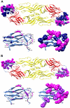
Figure 4. Location of epitope residues on E protein recognized by potent neutralizing mAbs.
Epitope residues of mAbs against DENV1 (A) and DENV2 (B). Epitope residues are highlighted with dark blue (DENV1 residues), purple (DENV2 residues), or magenta (DENV1 and DENV2 residues at the same position). Top view of E-E dimers (upper) and side view of domain III (lower right) with ribbon presentation of ß-strands and loops (lower left) are shown by the program UCSF chimera.

References

    1. Gubler DJ. Epidemic dengue/dengue hemorrhagic fever as a public health, social and economic problem in the 21st century. Trends Microbiol. 2002;10:100–103. - PubMed
    1. World Health Organization. Dengue hemorrhagic fever: Diagnosis, treatment, prevention and control, 3rd ed. Geneva, Switzerland: 2009.
    1. Whitehead SS, Blaney JE, Durbin AP, Murphy BR. Prospects for a dengue virus vaccine. Nat Rev Microbiol. 2007;5:518–528. - PubMed
    1. Lindenbach BD, Thiel HJ, Rice CM. Flaviviridae: the viruses and their replication, In: Knipe DM, Howley PM, editors. Fields virology, 5th ed. Philadelphia: Lippincott William & Wilkins; 2007. pp. 1101–1152.
    1. Heinz FX, Stiasny K. Flavivirus membrane fusion. J Gen Virol. 2006;87:2755–2766. - PubMed

Publication types

MeSH terms