. 2012 Mar;24(3):e113-24.
doi: 10.1111/j.1365-2982.2011.01860.x. Epub 2012 Jan 11.

Plasticity of mouse enteric synapses mediated through endocannabinoid and purinergic signaling

Affiliations

Plasticity of mouse enteric synapses mediated through endocannabinoid and purinergic signaling

I M Hons et al. Neurogastroenterol Motil. 2012 Mar.

Abstract

Background: The enteric nervous system (ENS) possesses extensive synaptic connections which integrate information and provide appropriate outputs to coordinate the activity of the gastrointestinal tract. The regulation of enteric synapses is not well understood. Cannabinoid (CB)(1) receptors inhibit the release of acetylcholine (ACh) in the ENS, but their role in the synapse is not understood. We tested the hypothesis that enteric CB(1) receptors provide inhibitory control of excitatory neurotransmission in the ENS.

Methods: Intracellular microelectrode recordings were obtained from mouse myenteric plexus neurons. Interganglionic fibers were stimulated with a concentric stimulating electrode to elicit synaptic events on to the recorded neuron. Differences between spontaneous and evoked fast synaptic transmission was examined within preparations from CB(1) deficient mice (CB(1)(-/-)) and wild-type (WT) littermate controls.

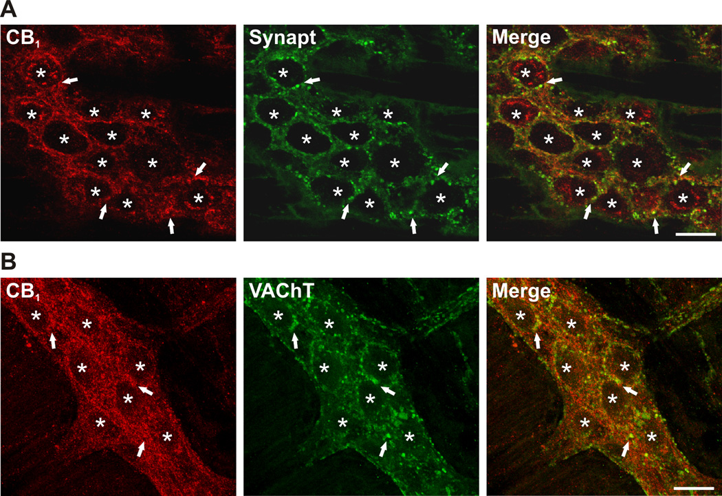
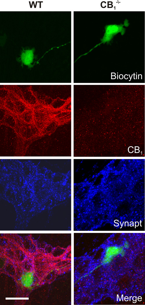
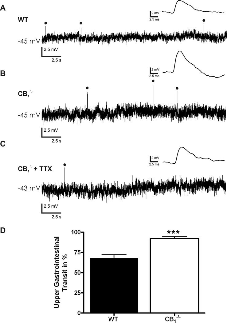
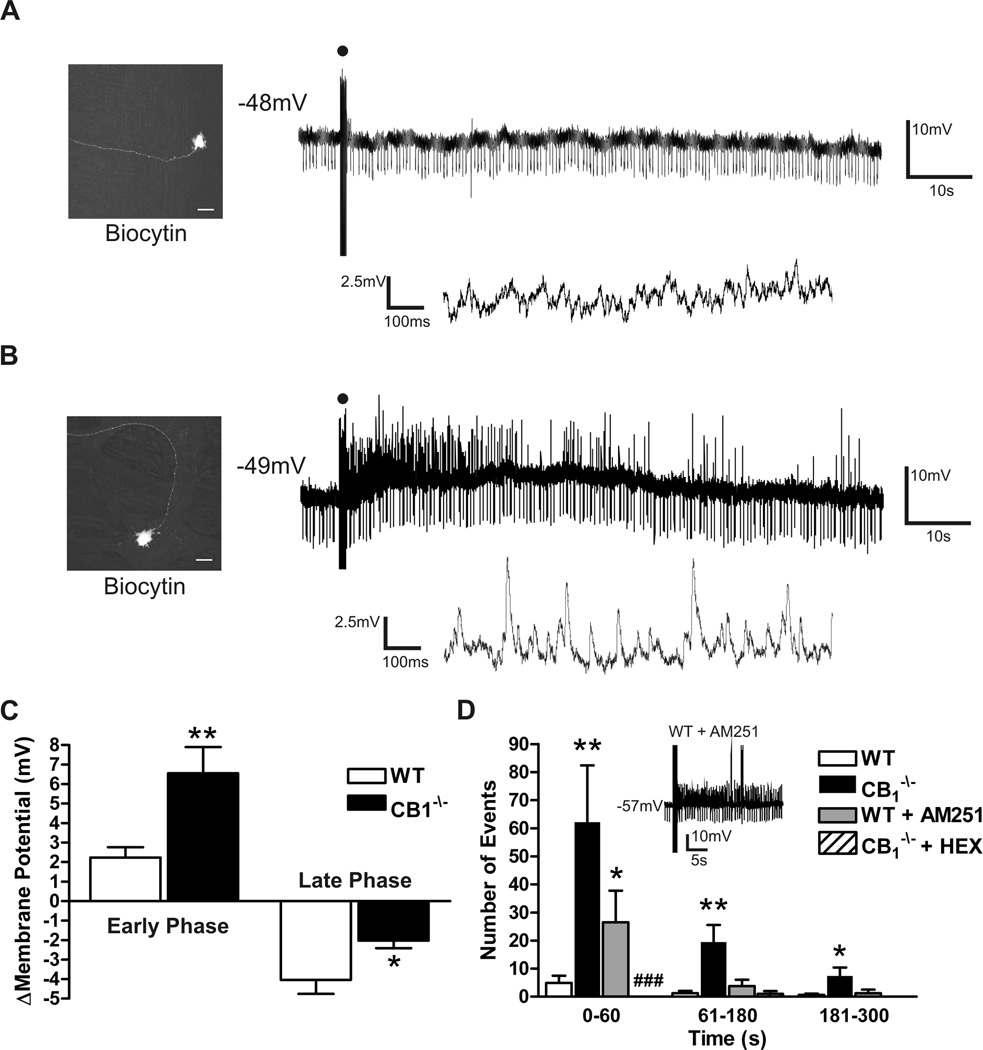
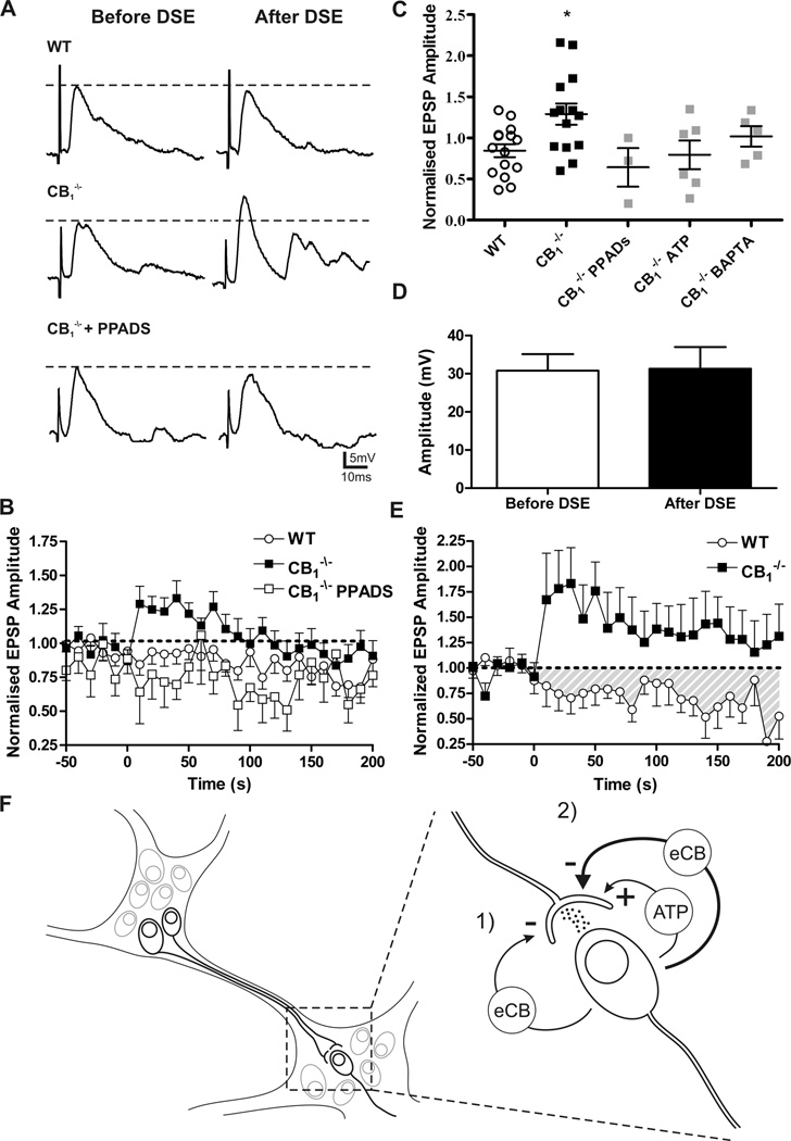
Key results: Cannabinoid receptors were colocalized on terminals expressing the vesicular ACh transporter and the synaptic protein synaptotagmin. A greater proportion of CB(1)(-/-) neurons received spontaneous fast excitatory postsynaptic potentials than neurons from WT preparations. The CB(1) agonist WIN55,212 depressed WT synapses without any effect on CB(1)(-/-) synapses. Synaptic activity in response to depolarization was markedly enhanced at CB(1)(-/-) synapses and after treatment with a CB(1) antagonist in WT preparations. Activity-dependent liberation of a retrograde purine messenger was demonstrated to facilitate synaptic transmission in CB(1)(-/-) mice.

Conclusions & inferences: Cannabinoid receptors inhibit transmitter release at enteric synapses and depress synaptic strength basally and in an activity-dependent manner. These actions help explain accelerated intestinal transit observed in the absence of CB(1) receptors.


Figures

Figure 1
Figure 1
Colocalization of the CB1 receptor with synaptotagmin (Synapt) and the vesicular ACh transporter (VAChT) in WT mouse ileal myenteric plexus. (A) Apparent colocalization of CB1 (red) occurs with Synapt (green) in the myenteric plexus. (B) A subpopulation of synapses are VAChT immunoreactive (green) and these synaptic terminals also appear to colocalize with the CB1 receptor (red). Representative confocal photomicrographs of localization studies performed in myenteric plexus preparations from 3 WT animals. Asterisks indicate the position of cell bodies within the enteric ganglion; arrows indicate areas of significant colocalization. Scale bars: 20 µm.
Figure 2
Figure 2
Spatial relationship between recorded neurons, CB1 receptor immunoreactivity and synaptic densities in myenteric S neurons. Colocalization of recorded neurons previously filled with biocytin (conjugated to FITC labelled avidin, green), with CB1 receptor (red) and synaptotagmin (Synapt, blue) immunoreactivity in WT and CB1−/− neurons. Scale bar: 20 µm.
Figure 3
Figure 3
Spontaneous synaptic events in CB1−/− and WT myenteric plexus preparations. (A) Spontaneous fast excitatory post-synaptic potentials (EPSPs) occurring in WT S neurons at resting membrane potential. (B) Spontaneous fast EPSPs occurring in CB1−/− S neurons at resting membrane potential. (C) Spontaneous fast EPSPs remain following incubation of CB1−/− preparations in 300nM tetrodotoxin (TTX) to block action potential conduction. (D) Significant increase in upper gastrointestinal transit in CB1−/− animals as indicated by an increased distance travelled by Evans blue given by gastric gavage 15min previously. Individual fast EPSPs within traces A-C are expanded and shown above the original recording. Spontaneously occurring fast EPSPs are indicated with closed circles. Resting membrane potential is indicated to the left of each recording. *** = P < 0.0001.
Figure 4
Figure 4
Demonstration of presynaptic action of CB1 receptors. Normalized first EPSP amplitude and the paired-pulse ratio PPR (30 ms interval) before and 4.5 minutes after the perfusion of 100 nM WIN55,212-2 on to WT neurons. In the presence of WIN55,212-2 the PPR was increased and first EPSP amplitude was reduced. ** P < 0.01.
Figure 5
Figure 5
Slow EPSPs evoked in WT and CB1−/− S neurons. (A) Lack of Slow EPSP or fast EPSPs in a WT myenteric S neuron. Lower trace indicates membrane potential activity 20 sec following the slow EPSP stimulus with increased time resolution. Slow membrane hyperpolarization typically occurs at a later time point, outside the sample traces. (B) Slow EPSP evoked in a CB1−/− S neuron demonstrating increased fast synaptic events as shown in the lower trace. (C) Maximum depolarization occurring within the first minute of the response (early phase) and maximum hyperpolarization during 5 min of recorded membrane potential dynamics (late phase) following slow EPSP stimulus. (D) Significant increase in fast synaptic events in CB1−/− S neurons can be mimicked in WT neurons with incubation of the tissue with the CB1 receptor antagonist AM251 (100 nM, sample trace d inset. Sample trace includes fast EPSP and APs). The fast synaptic events are entirely cholinergic as they are abolished by the addition of hexamethonium (HEX, 100µM). The proportion of neurons which received fast synaptic events during slow EPSP stimulus is significantly reduced with HEX, ### P < 0.0001 Chi squared test compared to control). * P < 0.05, ** P < 0.01 compared to control, Student’s unpaired T test. Micrographs in A and B demonstrate representative S neuron morphology of recorded neurons from WT and CB1−/− myenteric plexus. Scale bar in A and B: 20 µm. Downward deflections on membrane traces reflect current injections utilized to monitor input resistance.
Figure 6
Figure 6
Depolarization induced facilitation of fast EPSPs in CB1−/− S neurons. (A) Representative traces of evoked fast EPSP before and immediately after the postsynaptic depolarization protocol. There is no change in fast EPSP amplitude in WT neurons (upper), but there is a significant potentiation in CB1−/− (middle) neurons that is abolished in the presence of PPADs (lower). (B) Time course of the fast EPSP amplitude facilitation observed following the postsynaptic depolarization protocol. (C) Degree of fast EPSP facilitation following the postsynaptic stimulation protocol. Fast EPSPs are significantly potentiated in CB1−/− S neurons. This facilitation is blocked by PPADs, purine receptor desensitization, with 1 mM ATP (ATP) preincubation, and by intracellular calcium chelation of the postsynaptic neuron (BAPTA, 10mM). Groups were compared using one-way ANOVA with Dunnett’s post-test. (D) No change to postsynaptic sensitivity to ACh as demonstrated by the depolarization caused by picospritz application of 1mM ACh before and following the postsynaptic depolarization protocol. (E) Synaptic depression after coincidence stimulation in WT but not CB1−/− S neurons. The same experiment in CB1−/− S neurons caused significant facilitation. (F) Schematic representation of the organization and interconnectivity of ENS as well as synaptic regulation. 1) There is a tonic depression of synapses caused by endocannabinoids (eCB) acting through CB1. 2) High frequency postsynaptic depolarization induces the release of a purine (here ATP is shown) which potentiates further synaptic transmission. When this postsynaptic activity is paired with presynaptic activity there is a potentiation of eCB production and synaptic depression. * P < 0.05.

Similar articles

Cited by

References

    1. Furness JB. The enteric nervous system. Malden, Mass: Blackwell Pub; 2006.
    1. Galligan JJ, North RA. Pharmacology and function of nicotinic acetylcholine and P2X receptors in the enteric nervous system. Neurogastroenterol Motil. 2004;16 Suppl 1:64–70. - PubMed
    1. Lomax AE, Sharkey KA, Furness JB. The participation of the sympathetic innervation of the gastrointestinal tract in disease states. Neurogastroenterol Motil. 2010;22:7–18. - PubMed
    1. Wilson RI, Nicoll RA. Endogenous cannabinoids mediate retrograde signalling at hippocampal synapses. Nature. 2001;410:588–592. - PubMed
    1. Kreitzer AC, Regehr WG. Retrograde inhibition of presynaptic calcium influx by endogenous cannabinoids at excitatory synapses onto Purkinje cells. Neuron. 2001;29:717–727. - PubMed

Publication types

MeSH terms