. 2012 Mar;10(1):44-56.
doi: 10.1111/j.1476-5829.2011.00274.x. Epub 2011 May 18.

Bisphosphonates significantly increase the activity of doxorubicin or vincristine against canine malignant histiocytosis cells

Affiliations

Bisphosphonates significantly increase the activity of doxorubicin or vincristine against canine malignant histiocytosis cells

S D Hafeman et al. Vet Comp Oncol. 2012 Mar.

Abstract

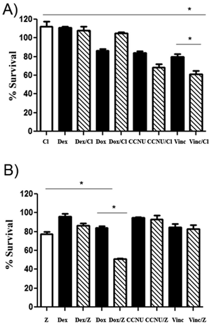
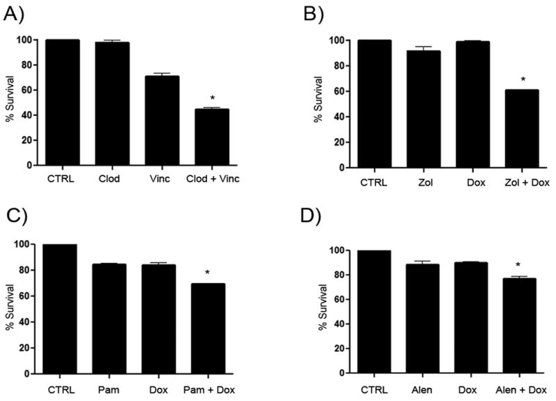
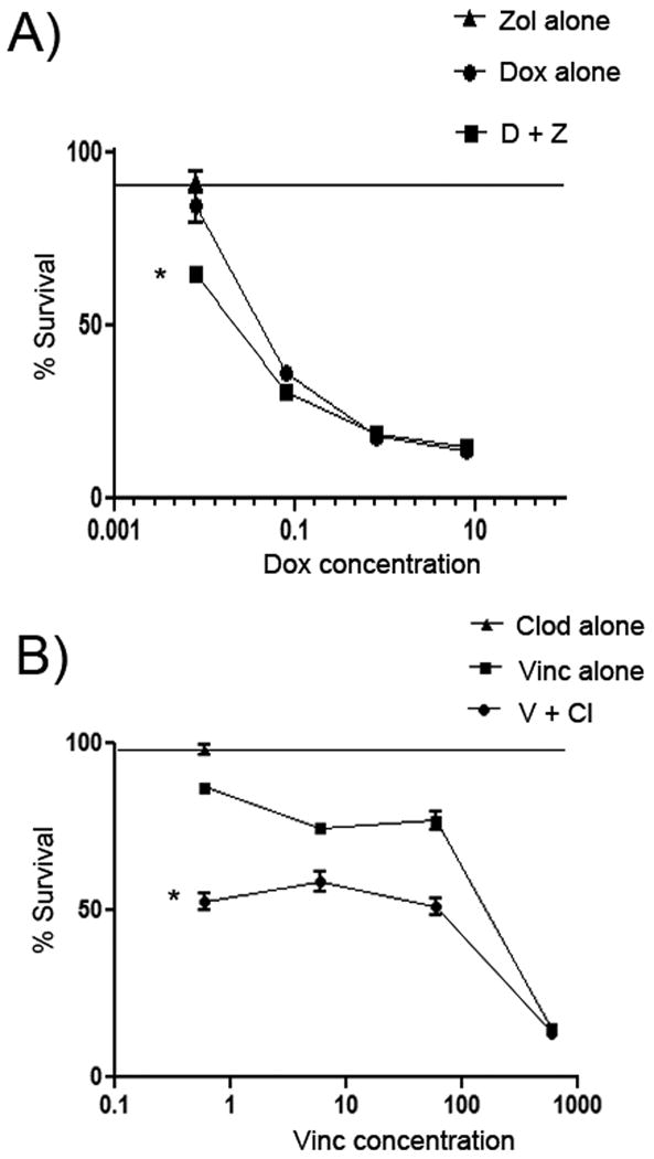
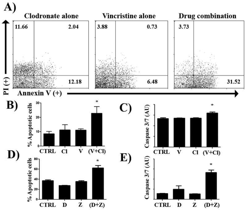
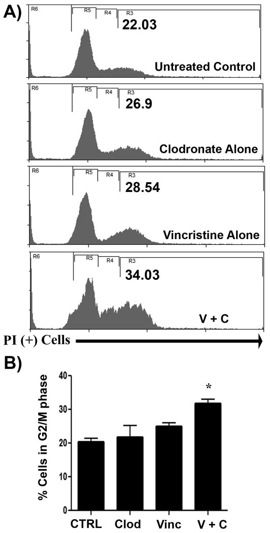
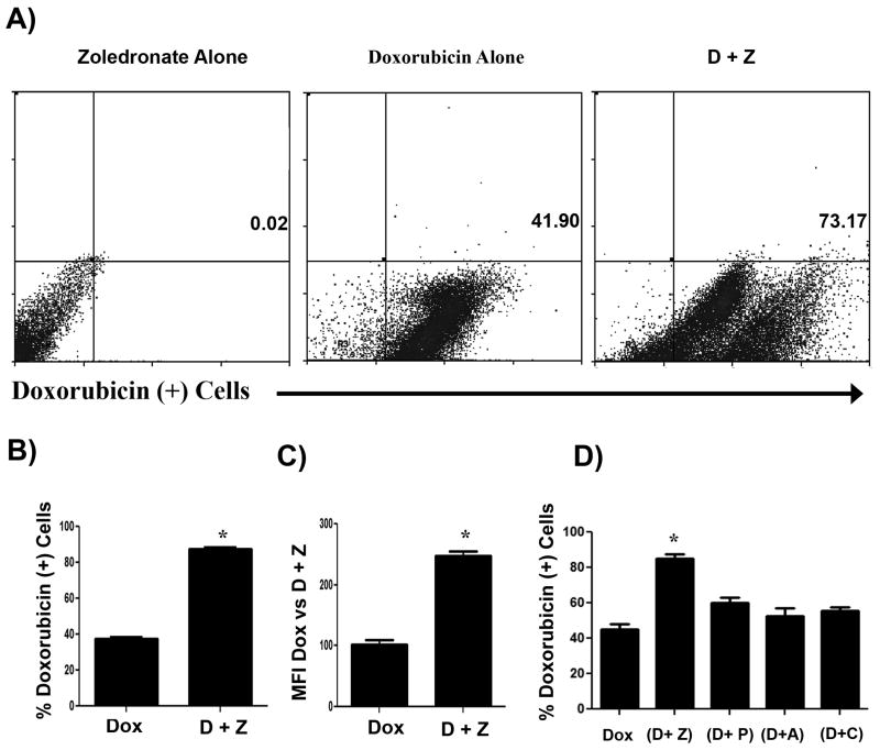
Canine malignant histiocytosis (MH) is an aggressive neoplasm of macrophages and dendritic cells. It carries a poor prognosis because of the development of widespread metastasis and poor sensitivity to chemotherapy. Thus, there is a large need for new treatments for MH. We hypothesized that bisphosphonates might be useful to increase the effectiveness of cytotoxic chemotherapy against MH. To address this question, we conducted in vitro screening studies using MH cell lines and a panel of 6 chemotherapy and 5 bisphosphonate drugs. The combination of clodronate with vincristine was found to elicit synergistic killing which was associated with a significant increase in cell cycle arrest. Second, zoledronate combined with doxorubicin also significantly increased cell killing. Zoledronate significantly increased the uptake of doxorubicin by MH cells. On the basis of these findings, we conclude that certain bisphosphonate drugs may increase the overall effectiveness of chemotherapy for MH in dogs.


Figures

Figure 1
Figure 1. Certain bisphosphonate and chemotherapy combinations elicit significantly increased killing of canine MH cells in vitro
In (A), the effects of clodronate on chemotherapy-induced killing of canine DH82 MH cells was assessed, using an MTT assay to assess tumor cell viability. Cells were treated with chemotherapy drug alone (black), clodronate alone (white), or with the combination of clodronate and chemotherapy drug (cross-hatch). In these assays, only the combination of clodronate and vincristine demonstrated a significant positive interaction (* = p < 0.05), as assessed by 1 way ANOVA with Tukey's post test. In (B), the effects of zoledronate on DH82 MH cell sensitivity to killing with chemotherapy drugs were assessed, using a similar approach as for (A). Cells were treated with chemotherapy drugs alone (black), zoledronate alone (white), or the two in combination (cross-hatch). The combination of zoledronate with doxorubicin showed a significant positive interaction (* = p < 0.05), as assessed by 1 way ANOVA with Tukey's post test. Cl = clodronate (5 μg/mL), Dex = dexamethasone (15 μg/mL), Dox = doxorubicin (0.2 μg/mL), CCNU = lomustine (1.5 μg/mL), vinc = vincristine (0.25 μg/mL), z = zoledronate (0.2 μg/mL).
Figure 2
Figure 2. Synergistic enhancement of MH cell killing by combinations of bisphosphonates with vincristine or doxorubicin
Significant killing of MH cells was seen in A-D when bisphosphonates were combined with chemotherapy as determined via MTT assay (A), DH82 MH cells were either untreated (CTRL) or treated with clodronate (Clod) or vincristine (Vinc) or both in combination (Clod + Vinc) for 72 hours. In (B), the interaction between zoledronate (Zol) and doxorubicin (Dox) was assessed. Similar experiments were done in (C) for the combination of pamidronate (Pam) with doxorubicin and in (D) for the combination of alendronate (Alen) and doxorubicin. (* = p < 0.05).
Figure 3
Figure 3. Dose response curves for determination of drug interactions between zoledronate and doxorubicin or between clodronate and vincristine
In (A), dose response curves were generated for DH82 cells treated with zoledronate, doxorubicin, or zoledronate plus doxorubicin in order to compare drug interactions via Bliss analysis. Cell viability was significantly reduced in cells treated with zoledronate (0.2 μg/mL) and increasing doses of doxorubicin, compared to cells treated with zoledronate alone (0.2 μg/mL) or increasing doses of doxorubicin alone, as assessed by Bliss analysis. In (B), a similar analysis was conducted using DH82 cells treated with clodronate (5 μg/mL) alone, with clodronate (5 μg/mL) plus increasing doses of vincristine, or with increasing doses of vincristine alone. The viability of MH cells was found using Bliss analysis to be significantly reduced in cells treated with the combination of clodronate and vincristine. (* = p < 0.05)
Figure 4
Figure 4. Combined treatment with zoledronate and doxorubicin and clodronate and vincristine results in increased MH cell apoptosis
DH82 cells were treated with clodronate (5 μg/mL) and vincristine (0.25 μg/mL), alone or in combination, for 48 hours and cell apoptosis was assessed using Annexin V and propidium iodide staining, as described in Methods. In (A), an increased percentage of Annexin V+ cells were noted in cells treated with the combination of two drugs, as assessed by flow cytometry and depicted in these representative FACS plots. In (B), the mean percentage of apoptotic cells was compared between MH cells treated with clodronate or vincristine alone or in combination. There was a significant increase (* = p < 0.05) in the percentage of apoptotic cells in the combination treated group, as assessed by ANOVA and Tukey's multiple means comparison test. In (C), induction of apoptosis was also assessed by measuring induction of caspase 3 and 7 activity, as described in Methods. Treatment of DH82 cells with the combination of clodronate (5 μg/mL), vincristine (0.25 μg/mL), or both induced a significant increase (*= p < 0.05) in caspase 3/7 fluorescence (AU, arbitrary fluorescence units) as compared to untreated control cells or cells treated with one drug only, as assessed by ANOVA. In (D), combined treatment with zoledronate (0.2 μg/mL) and doxorubicin (0.2 μg/mL) induced a significant increase in the number of apoptotic MH cells, as assessed by Annexin V and propidium iodide staining. In (E), the combined treatment with zoledronate (0.2 μg/mL) and doxorubicin (0.2 μg/mL) resulted in a significant (p < 0.05) increase in caspase 3 and 7 activity in MH cells, compared to cells treated with either drug alone, as assessed by ANOVA. Each of the experiments depicted were repeated independently two additional times and similar results were obtained.
Figure 5
Figure 5. Addition of clodronate to vincristine treatment increased cell cycle arrest in canine MH cells
The effect of the addition of clodronate to vincristine-treated DH82 cells was assessed using flow cytometry and cell cycle analysis. In (A), representative flow cytometry histograms are displayed for cells treated for 48 hours with clodronate (5 μg/mL), vincristine (0.2 μg/mL), or with both in combination. In cells treated with the combination of both drugs, there as a notable increase in the percentage of DH82 cells exhibiting G2/M arrest (R3 = G2/M). In (B), the mean percentages of cells in the G2/M stage of cell cycle progression were calculated for untreated cells or cells treated with clodronate or vincristine, alone or together. A significant increase (* = p < 0.05) in cells in the G2/M stage was observed in cells treated with clodronate plus vincristine, as assessed by ANOVA.
Figure 6
Figure 6. Treatment with zoledronate increases doxorubicin uptake by DH82 cells
DH82 cells were treated with doxorubicin alone (0.2 μg/mL), or with zoledronate (0.2 μg/mL) plus doxorubicin for 24 hours. The cells were then analyzed for intracellular concentrations of doxorubicin using flow cytometry. In (A), representative dot plots of doxorubicin fluorescence intensity for cells treated with zoledronate alone, doxorubicin alone, or zoledronate plus doxorubicin are depicted. In (B), the mean percentage of doxorubicin+ MH cells from triplicate wells treated with doxorubicin alone or doxorubicin plus zoledronate was plotted. The percentage of doxorubicin+ cells was significantly increased (* = p < 0.05) in the combination treated cells. In (C), the mean of the mean fluorescence intensities (MFI) of doxorubicin expression by cells treated with doxorubicin alone or doxorubicin plus zoledronate was plotted. The mean MFI of doxorubicin expression was significantly increased (* = p < 0.05) in the combination treated cells. In (D), MH cells in triplicate wells were treated doxorubicin alone or with zoledronate (Z), pamidronate (P), alendronate (A), or clodronate (C) plus doxorubicin for 24 hours and the mean percentage of doxorubicin+ cells was determined by flow cytometry. Only treatment with zoledronate produced a significant increase (* = p < 0.05) in the percentage of doxorubicin+ cells, as assessed by ANOVA.

Similar articles

Cited by

References

    1. Affolter VK, Moore PF. Canine cutaneous and systemic histiocytosis: reactive histiocytosis of dermal dendritic cells. The American Journal of dermatopathology. 2000;22(1):40–8. - PubMed
    1. MacEwen EG, Withrow SJ. Small Animal Clinical Oncology. Philadelphia PA: Saunders Co; 1996.
    1. Fulmer AK, Mauldin GE. Canine histiocytic neoplasia: an overview. Can Vet J. 2007;8:1041–1043. 1046–1050. - PMC - PubMed
    1. Affolter VK, Moore PF. Localized and disseminated histiocytic sarcoma of dendritic cell origin in dogs. Veterinary pathology. 2002;39(1):74–83. - PubMed
    1. Moore PF, Affolter VK, Vernau W. Canine hemophagocytic histiocytic sarcoma: a proliferative disorder of CD11d+ macrophages. Veterinary pathology. 2006;43(5):632–45. - PubMed

Publication types

MeSH terms