

Validation of an LC-MS/MS method to determine five immunosuppressants with deuterated internal standards including MPA
- PMID: 22236286
- PMCID: PMC3398287
- DOI: 10.1186/1472-6904-12-2
Validation of an LC-MS/MS method to determine five immunosuppressants with deuterated internal standards including MPA
Abstract
Background: Therapeutic drug monitoring of immunosuppressive drugs in organ-transplanted patients is crucial to prevent intoxication or transplant rejection due to inadequate dosage. The commonly used immunoassays have been gradually undergoing replacement by mass spectrometry, since this physical method offers both a higher sensitivity and specificity. However, a switch should be carefully considered because it is a challenging procedure and needs to be thoroughly validated. From an economic perspective it is reasonable to include mycophenolic acid into the assay, because this saves the necessity for an additional measurement. However, to date very few validation protocols for the measurement of immunosuppressants, including mycophenolic acid, are available. In order to adequately compensate for matrix effects, the use of stable isotope labeled internal standards is advisable. Here, the authors describe a single method suitable for the quantification of cyclosporine A, tacrolimus, sirolimus, everolimus and mycophenolic acid, based on deuterated internal standards.
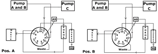
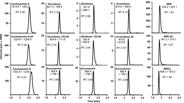
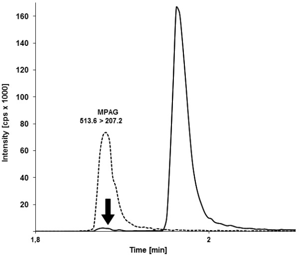
Methods: Plasma proteins were precipitated with zinc-sulfate, followed by an online solid phase extraction in the flow-through direction. Chromatographic separation was performed by a c18-phenyl-hexyl column. For subsequent mass spectrometric analysis stable-isotope-labeled internal standards were used. Results were available after 3.5 minutes.
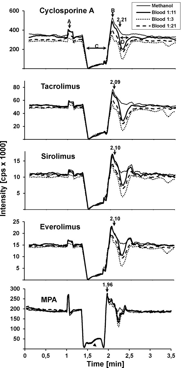
Results: Low quantification limits (accuracy: 104 - 118%) and linearity resulted in 2 -1250 ng/ml for cyclosporine A; 0.5 - 42.2 ng/ml for tacrolimus; 0.6 - 49.2 ng/ml for sirolimus; 0.5 - 40.8 ng/ml for everolimus and 0.01 - 7.5 μg/ml for mycophenolic acid. Intra-assay precision revealed a coefficient of variation (CV) of 0.9 - 14.7%, with an accuracy of 89 - 138%. The CV of inter-assay precision was 2.5 - 12.5%, with an accuracy of 90 - 113%. Recovery ranged from 76.6 to 84%. Matrix effects were well compensated by deuterated internal standards.
Conclusions: The authors present a fast, economical and robust method for routine therapeutic drug monitoring comprising five immunosuppressants including mycophenolic acid.
Figures
References
-
- Beckebaum S, Armstrong VW, Cicinnati VR, Streit F, Klein CG, Gerken G, Paul A, Oellerich M. Pharmacokinetics of mycophenolic acid and its glucuronide metabolites in stable adult liver transplant recipients with renal dysfunction on a low-dose calcineurin inhibitor regimen and mycophenolate mofetil. Ther Drug Monit. 2009;31(2):205–210. doi: 10.1097/FTD.0b013e31819743d9. - DOI - PubMed
-
- Schneider HSW. Tandem-Massenspektrometrie in der therapeutischen Arzneimittelspiegel-Bestimmung: Praxis und Tücken der Anwendung. J Lab Med. 2006;6(30):428–437.
Publication types
MeSH terms
Substances
LinkOut - more resources
Full Text Sources
Other Literature Sources
