. 2012;79(1):34-44.
doi: 10.1159/000332215. Epub 2012 Jan 12.

Immunolocalization of sprouty-1 and sprouty-2 in developing rat lung

Affiliations

Immunolocalization of sprouty-1 and sprouty-2 in developing rat lung

Shuichi Hashimoto et al. Pathobiology. 2012.

Abstract

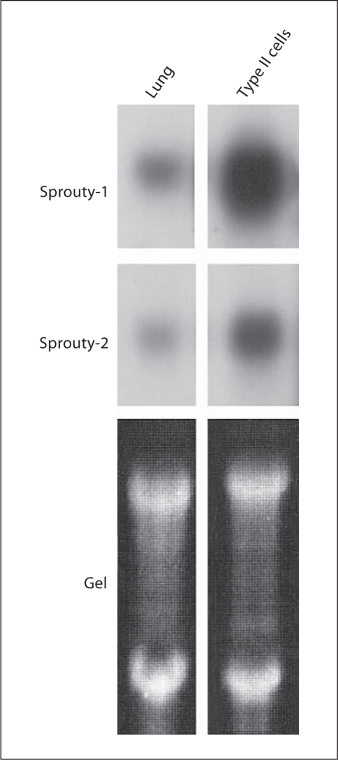
Objective: Sprouty, a common antagonist of fibroblast growth factor (FGF) and epidermal growth factor signaling, is a key player regulating tracheal branching and eye development in Drosophila. Four Sprouty homologs have been identified in vertebrates and all share a cysteine-rich region. However, the physiological function(s) of the individual Sprouty homologs is unknown. mRNA of Sprouty homologs is expressed during mouse lung development. In the present study, we investigated the immunolocalization of Sprouty proteins in rat lung at different stages of development.

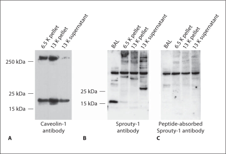
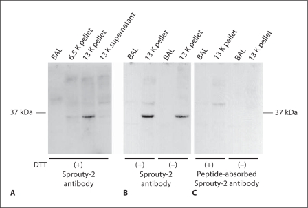
Methods: Rabbit antibodies were raised against peptides derived from rat Sprouty-1 and Sprouty-2 and were used in Western blot analysis to determine Sprouty distribution in subcellular fractions (pellets and supernatant centrifuged at 5,000 and 20,000 g) and bronchoalveolar lavage fluid (BAL) from adult rat lungs or used in immunohistochemistry.

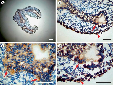
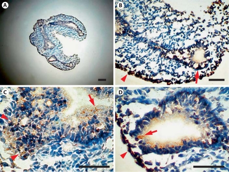
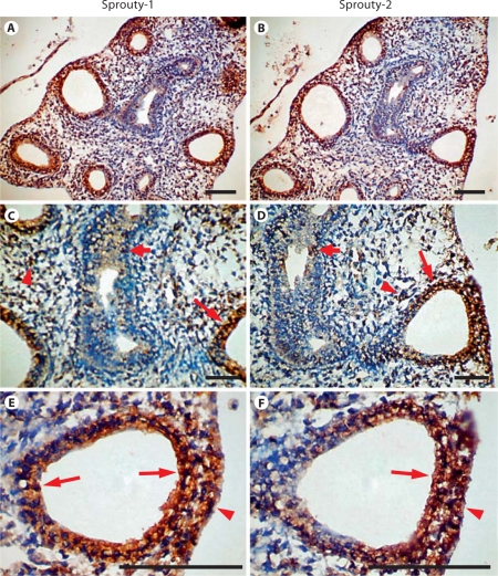
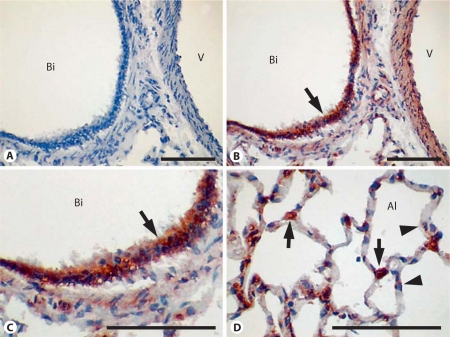
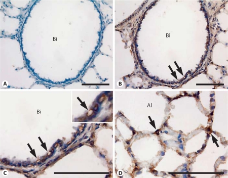
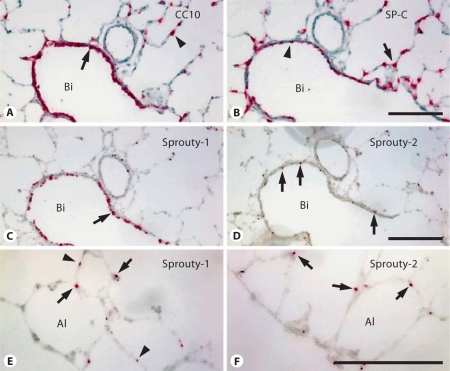
Results: Western blot analysis revealed a 30-kDa Sprouty-1 band and a 34-kDa Sprouty-2 band in the supernatant and pellet fractions centrifuged at 20,000 g. BAL contained a band of approximately 16 kDa with Sprouty-1 antibody derived from proteolytic fragmentation of Sprouty-1. In embryonic day (E) 14 and E16 lungs, Sprouty-1 and Sprouty-2 were expressed both in epithelial and peripheral mesenchymal cells. In adult rat lung, bronchiolar and alveolar type II epithelial cells showed staining for both Sprouty-1 and Sprouty-2. Sprouty-1 expression was also seen in alveolar type I epithelial cells.

Conclusion: In light of the proximity of the distribution of Sprouty to that of FGF-10 (peripheral mesenchyme) and its receptor FGFR2IIIb (distal tubular epithelium) in lung development, and the finding that FGF-9, which is expressed in mesothelial cells, upregulates FGF-10, it appears that Sprouty expression in epithelial and mesenchymal cells during branching morphogenesis is closely related to signaling by FGF-9 and FGF-10.


Figures

Fig. 1
Fig. 1
Characterization of Sprouty-1 antibody by Western blot analysis of lung subcellular fractions. Three fractions (6.5 K pellet, 13 K pellet, membrane-rich and 13 K supernatant) were obtained by differential centrifugation of homogenates of adult rat lungs. An additional fraction was obtained by concentrating BAL obtained from adult rat lungs. A Each fraction obtained by differential centrifugation shows the presence of caveolin-1. The highest amount of caveolin was seen in the 13 K pellet fraction. These fractions were further used for characterization of Sprouty-1 polyclonal antibody by Western blot analysis. B Blot treated with Sprouty-1 antibody. C Blot treated with peptide-absorbed Sprouty-1 antibody. Specific bands of approximately 16 and 30 kDa were detected in the BAL and 13 K supernatant fractions, respectively (B). These bands were not detectable using preabsorbed Sprouty-1 antibody (C).
Fig. 2
Fig. 2
Characterization of Sprouty-2 antibody by Western blot analysis of lung subcellular fractions. The fractions described in figure 1 were also used for characterization of Sprouty-2 antibody by Western blot analysis. A, B Blot treated with Sprouty-2 antibody. C Blot treated with peptideabsorbed Sprouty-2 antibody. A specific band of approximately 34 kDa was detected in the 13 K pellet fraction. This band was not detectable using preabsorbed Sprouty-2 antibody (C). No specific band was detected in the BAL or 13 K supernatant fractions (A).
Fig. 3
Fig. 3
Northern blot analysis of Sprouty-1 and Sprouty-2 mRNA expression in adult rat lung and isolated alveolar epithelial type II cells. High expression of mRNA of both Sproutys is seen in the lung and type II cells. The latter appear to be the major cell type expressing Sproutys in the adult rat lung.
Fig. 4
Fig. 4
Expression of Sprouty-1 in lung at the early pseudoglandular stage (E14). Sections from lungs of E14 rat embryos were immunostained with Sprouty-1 antibody (A). Staining for Sprouty-1 is seen at the basolateral surfaces of epithelial cells (arrows in B, D) and in mesenchymal cells adjacent to airway epithelium (arrows in C) and at the periphery (arrowheads in B, D). Staining is especially strong in clusters of epithelial cells at the leading edges of branching airways. Bar = 100 μm.
Fig. 5
Fig. 5
Expression of Sprouty-2 in lung at the early pseudoglandular stage (E14). Sections from lungs of E14 rat embryos were immunostained with Sprouty-2 antibody (A). Staining for Sprouty-2 is seen at the lumenal surfaces of epithelial cells lining tubules (arrows in C, D) and in clusters of epithelial cells at the leading edges of branching tubules (arrow in B, arrowheads in C). Peripheral mesenchymal cells are also stained (arrowheads in B, D). Bar = 100 μm.
Fig. 6
Fig. 6
Expression of Sprouty-1 and Sprouty-2 during the midpseudoglandular stage of lung development (E16). Sections from lungs of E16 rat embryos were immunostained with Sprouty-1 antibody (A, C, E) or Sprouty-2 antibody (B, D, F). Staining for Sprouty-1 and Sprouty-2 is seen in the epithelium of distal tubules (long arrows in C–F) as well as in the peripheral mesenchymal cells (arrowheads in E, F). While all epithelial cells of distal tubules are uniformly stained for Sprouty-1 (arrows in E), staining for Sprouty-2 is limited to the epithelial cells closer to the periphery of the lung (arrow in F). Low levels of Sprouty-1 and Sprouty-2 were detected in mesenchyme (arrowheads in C, D) and proximal airway epithelium (short arrows in C, D). Bar = 100 μm.
Fig. 7
Fig. 7
Sprouty-1 and Sprouty-2 expression in E18 lung. Sections of E18 lungs were immunostained using Sprouty-1 (A, C) and Sprouty-2 (B, D) antibodies. Strong staining for both Sproutys is seen in the distal epithelium (arrows in A–D). Expression of these proteins is weak in the proximal epithelium (arrowheads in A, B). Peripheral mesenchymal staining is barely detected at this stage of lung development (arrowheads in C, D). Bi = Bronchiole; Al = alveolus. Bar = 100 μm.
Fig. 8
Fig. 8
Dual-probe in situ hybridization to monitor the expression of SP-C, Sprouty-1 and Sprouty-2 mRNA in E18 lung. A Overlapping expression of Sprouty-1 (red; colors only in online version) and SP-C (brown) mRNA was observed in epithelial cells of the distal alveolar buds (arrows), whereas only Sprouty-1 mRNA expression was observed in the peribronchiolar mesenchymal cells (arrowheads). B Overlapping expression of Sprouty-2 (red) and SP-C (brown) mRNA is also seen in the epithelial cells of the distal alveolar buds (arrows). Sprouty-2 mRNA expression is not seen in peribronchiolar mesenchymal cells (arrowhead). C, D Overall distribution of Sprouty-1 (brown) and Sprouty-2 (red) mRNA is overlapping in distal alveolar buds (arrows in C, D) and distinct in peribronchiolar mesenchymal cells with regard to Sprouty-1 mRNA expression (arrowheads in C). Bi = Bronchiole; Al = alveolus. Bar = 100 μm.
Fig. 9
Fig. 9
Sprouty-1 expression in adult rat lung. Sections of adult rat lung were immunostained using Sprouty-1 antibody. A Treatment with peptide-absorbed Sprouty-1 antibody. B, C Bronchiolar epithelium at different magnifications. D Alveolar epithelium. Bronchial epithelium shows strong staining (arrows in B, C). In the alveolar region, type II (arrows in D) and type I epithelial cells (arrowheads in D) are frequently stained. There is no staining in the section treated with preabsorbed antibody (A). Bi = Bronchiole; Al = alveolus; V = vessel. Bar = 100 μm.
Fig. 10
Fig. 10
Sprouty-2 expression in adult rat lung. Sections of adult rat lung were immunostained using Sprouty-2 antibody. A Treatment with peptide-absorbed Sprouty-2 antibody. B, C Bronchiolar epithelium at different magnifications. D Alveolar epithelium. Intense staining is seen in bronchial epithelial cells. In these cells, the membranes surrounding the intracellular vacuoles/vesicles are also stained (arrows in B, C and inset). In the alveolar region, type II epithelial cells are frequently stained (arrows in D). No staining is seen in sections treated with preabsorbed antibody (A). Bi = Bronchiole; Al = Alveolus. Bar = 100 μm.
Fig. 11
Fig. 11
In situ hybridization to compare the expression of Sprouty-1, Sprouty-2, CC10 and SP-C mRNA in the adult rat lung. A Bronchiolar epithelial cells show high CC10 mRNA expression (arrow). CC10 mRNA expression is also seen in a smaller number of alveolar epithelial type II cells (arrowhead). B In contrast, a distinct distribution of SP-C mRNA in alveolar epithelial type II cells was observed (arrow), with expression in a smaller number of bronchiolar epithelial cells (arrowhead). C, D Sprouty-1 and, to a lesser extent, Sprouty-2 mRNA are expressed in the bronchiolar epithelial cells (arrows). E, F In the alveolar region, expression of both Sproutys is seen in type II cells (arrows). Sprouty-1 mRNA is also expressed in type I cells (arrowheads in E). Bi = Bronchiole; Al = alveolus. Bar = 100 μm.

Similar articles

Cited by

References

    1. Metzger RJ, Klein OD, Martin GR, Krasnow MA. The branching programme of mouse lung development. Nature. 2008;453:745–750. - PMC - PubMed
    1. Morrisey EE, Hogan BL. Preparing for the first breath: genetic and cellular mechanisms in lung development. Dev Cell. 2010;18:8–23. - PMC - PubMed
    1. Casci T, Vinos J, Freeman M. Sprouty, an intracellular inhibitor of ras signaling. Cell. 1999;96:655–665. - PubMed
    1. Hacohen N, Kramer S, Sutherland D, Hiromi Y, Krasnow MA. Sprouty encodes a novel antagonist of FGF signaling that patterns apical branching of the Drosophila airways. Cell. 1998;92:253–263. - PubMed
    1. Kramer S, Okabe M, Hacohen N, Krasnow MA, Hiromi Y. Sprouty: a common antagonist of FGF and EGF signaling pathways in Drosophila. Development. 1999;126:2515–2525. - PubMed

Publication types