. 2012 Apr;107(8):2072-82.
doi: 10.1152/jn.00730.2011. Epub 2012 Jan 11.

Somatosensory control of balance during locomotion in decerebrated cat

Affiliations

Somatosensory control of balance during locomotion in decerebrated cat

Pavel Musienko et al. J Neurophysiol. 2012 Apr.

Abstract

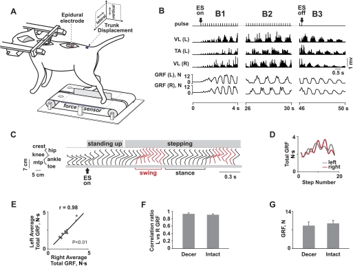
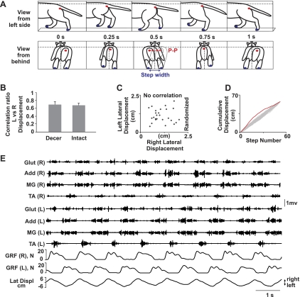
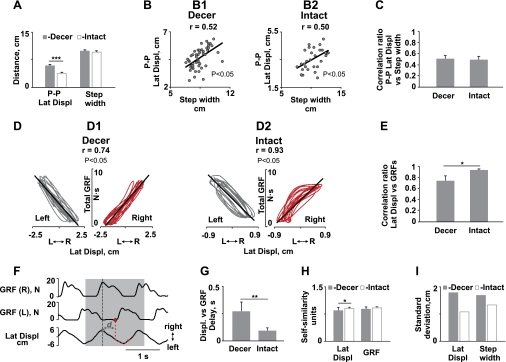
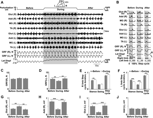
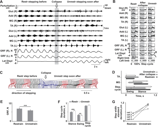
Postmammillary decerebrated cats can generate stepping on a moving treadmill belt when the brain stem or spinal cord is stimulated tonically and the hindquarters are supported both vertically and laterally. While adequate propulsion seems to be generated by the hindlimbs under these conditions, the ability to sustain equilibrium during locomotion has not been examined extensively. We found that tonic epidural spinal cord stimulation (5 Hz at L5) of decerebrated cats initiated and sustained unrestrained weight-bearing hindlimb stepping for extended periods. Detailed analyses of the relationships among hindlimb muscle EMG activity and trunk and limb kinematics and kinetics indicated that the motor circuitries in decerebrated cats actively maintain equilibrium during walking, similar to that observed in intact animals. Because of the suppression of vestibular, visual, and head-neck-trunk sensory input, balance-related adjustments relied entirely on the integration of somatosensory information arising from the moving hindquarters. In addition to dynamic balance control during unperturbed locomotion, sustained stepping could be reestablished rapidly after a collapse or stumble when the hindquarters switched from a restrained to an unrestrained condition. Deflecting the body by pulling the tail laterally induced adaptive modulations in the EMG activity, step cycle features, and left-right ground reaction forces that were sufficient to maintain lateral stability. Thus the brain stem-spinal cord circuitry of decerebrated cats in response to tonic spinal cord stimulation can control dynamic balance during locomotion using only somatosensory input.


Figures

Fig. 1.
Fig. 1.
Weight-bearing hindlimb stepping in decerebrated cats facilitated by epidural electrical stimulation (ES). A: cat secured in a stereotaxic frame. Stimulating electrode is sutured epidurally (at spinal level L5). Position sensors are attached to the pelvis, and displacement and force sensors are placed beneath each belt to record ground reaction forces (GRFs) from the right (R) and left (L) hindlimbs. B: R and L vastus lateralis (VL) and L tibialis anterior (TA) rectified EMGs and GRFs are shown during initiation of locomotor activity by ES (B1), during continuous stepping (B2), and after ES is turned off (B3). C: stick diagrams (60 ms between sticks) of joint movements after the initiation of ES during the transition from sitting to standing and for the initial step cycle [swing (red) and stance are indicated by brackets]; crest, iliac crest; mtp, metatarsophalangeal. D: sequence of R-L GRFs for consecutive steps showing a close relationship between left and right motor responses that gradually increase followed by a progressive decrease during a series of continuous steps. E: average correlation for the L and R total GRFs within the entire duration of the stepping trial for 10 experiments in decerebrated cats (n = 7 cats, P < 0.01). F: average correlation ratios for L vs. R GRFs plotted on a step-to-step basis during bipedal hindlimb stepping in decerebrated (Decer, n = 4 cats, 10–15 steps from each limb per cat) and intact (n = 4 cats, 10–15 steps from each limb per cat) animals. G: average amplitude of GRFs in decerebrated (mean ± SE, n = 4 cats, 10–15 steps from each limb per cat) and intact (n = 4 cats, 10–15 steps from each limb per cat) animals.
Fig. 2.
Fig. 2.
Kinematics, kinetics, and EMG patterns of the hindquarters dynamic destabilization during unrestrained hindlimb stepping in decerebrated cats. A: movement viewed from the L side and from behind. The hindquarters deviated vertically and laterally during stepping initiated by ES, with each successive step compensating for the previous step. Peak-to-peak (P-P) lateral displacements of the pelvis and the step width (distance between the stance paws in the frontal plane) are shown. B: averaged correlation ratios for L vs. R displacements of the trunk during bipedal hindlimb stepping in decerebrated (n = 4 cats, 10–15 steps per cat) and intact (n = 4 cats, 10–15 steps per cat) animals. C: there is no correlation when all R and L displacements are randomized with respect to their order of occurrence. D: cumulative R and L pelvis displacements plotted in order of occurrence (red line) or randomized (Monte Carlo 500 times, gray line). Note that the actual cumulative sequence quickly falls outside the probability that would be expected if the R-L limb sequences were random. E: R and L medial gastrocnemius (MG), tibialis anterior (TA), adductor femoris (Add), and gluteus medius (Glut) EMG activity, GRFs, and lateral trunk displacements (Lat Displ) of decerebrated cats during weight-bearing unrestrained locomotion.
Fig. 3.
Fig. 3.
Balance-related responses in decerebrated and intact cats. A: averaged values of P-P lateral displacements (Lat Displ) of the pelvis and step width in decerebrated (n = 4 cats, 9–15 steps per cat) and intact (n = 4 cats, 9–15 steps per cat) cats. B: correlation ratios for P-P lateral displacements vs. step width plotted on a step-to-step basis in decerebrated (B1) and intact (B2) cats. Graphs were created based on data from 1 cat (n = 48 steps for decerebrated, n = 35 steps for intact). C: comparison of the average correlation ratios for P-P lateral displacements vs. step width in decerebrated (n = 4 cats, 10–15 steps per cat) and intact (n = 4 cats, 10–15 steps per cat) cats. D: R-L correlations between GRFs and pelvis displacements in decerebrated (D1) and intact (D2) cats. Graphs were created based on data from 1 cat from each group (n = 10–15 steps). E: comparison of the average correlation ratios for lateral displacements vs. ipsilateral GRFs during bipedal hindlimb stepping in decerebrated (n = 4 cats, 10–15 steps from each limb per cat) and intact (n = 4 cats, 10–15 steps from each limb per cat) cats. F: delay (d) between the peak lateral displacement (black dashed lines) and the following GRF response. The vertical red dashed line in the gray shaded area marks the initiation of the increase in the GRF. G–I: average delay between lateral displacements (Displ) and the ipsilateral GRF (G), self-similarity coefficients in normalized units for lateral displacements and GRFs (H), and standard deviation of lateral displacements and step width (I) in decerebrated (n = 4 cats, 10–15 steps per cat) and intact (n = 4 cats, 10–15 steps per cat) animals. Significant difference between conditions: *P < 0.05, **P < 0.01, ***P < 0.001.
Fig. 4.
Fig. 4.
Adaptive postural responses to prolonged lateral perturbation during hindlimb stepping in decerebrated cats. A: EMG activity, GRFs, and lateral body displacements before, during, and after the pelvis is being perturbed laterally to the right (shaded areas). B: average (1 SE, shaded area) rectified EMG signals from the L and R Glut, Add, MG, and TA, L and R GRFs, and lateral pelvis displacements during the stance (St) and swing (Sw) phases of gait cycles before, during, and after perturbation for the raw data in A; 9–10 gait cycles were averaged for each period. C–F: average GRF magnitudes generated by L and R hindlimbs and the duration of the swing-stance phases of L and R hindlimb step cycles before, during, and after lateral perturbation for all tested animals (n = 4 cats, 10–15 steps from each limb per cat). G–J: bilateral modulation of EMG amplitudes [R Glut (G) and Add (H), L Glut (I) and Add (J)] after perturbation averaged from all experiments (n = 4, 10–15 steps from each limb per cat). Significant difference between conditions: **P < 0.01, ***P < 0.001.
Fig. 5.
Fig. 5.
Adaptive postural responses after collapse during hindlimb stepping in decerebrated cats. A: EMG activity, GRFs, and lateral pelvis displacements during stepping with restrained pelvis before collapse (Restr stepping before) from the clamps (shaded area) and unrestrained stepping soon after (Unrestr stepping soon after). B: average (1 SE, shaded area) rectified EMG signals from L and R Glut, Add, MG, and TA, L and R GRFs, and lateral pelvis displacements during the stance (St) and swing (Sw) phases of gait cycles during restrained stepping before collapse (Restr), unrestrained stepping soon after (After collapse), and stabilized unrestrained stepping (Unrestr) for the raw data in A from 1 cat; 9–10 gait cycles were averaged for each period. C: stick diagrams (50 ms between sticks, red lines represent the swing phase) of the joint movements for a sequence of stepping under the restrained condition and unrestrained stepping soon after the release of the pelvis clamp resulting in a collapse of the hindquarters and a stumble (blue). D: average duration of EMG bursts in the ipsilateral flexor (TA) and extensor (MG) muscles during the stance and swing phases of gait cycle before the collapse (Restrain), unrestrained stepping soon after (After collapse), and stable unrestrained stepping (Unrestrain); 7–10 gait cycles from 1 cat were averaged for each period. E and F: average GRF magnitudes generated by both the L and R hindlimbs and the duration of swing-stance phases of the step cycle in restrained and stable unrestrained stepping for 4 tested animals (n = 4 cats, 15–20 steps per cat). G: differences in standard deviation of the step width value during restrained compared with unrestrained locomotion (n = 15 steps per condition in 4 cats). Significant difference between conditions: **P < 0.01, ***P < 0.001.

References

    1. Armstrong DM. Supraspinal contributions to initiation and control of locomotion in the cat. Prog Neurobiol 26: 273–361, 1986 - PubMed
    1. Arshavsky YI, Gelfand IM, Orlovsky GN. The cerebellum and control of rhythmical movements. Trends Neurosci 6: 417–422, 1983
    1. Barbeau H, Rossignol S. Recovery of locomotion after chronic spinalization in the adult cat. Brain Res 412: 84–95, 1987 - PubMed
    1. Barberini CL, Macpherson JM. Effect of head position on postural orientation and equilibrium. Exp Brain Res 122: 175–184, 1998 - PubMed
    1. Bard P, Macht MB. The behavior of chronically decerebrate cats. In: Neurological Basis of Behavior, edited by Wolstenholme GEW, O'Connor CM. London: Churchill, 1958, p. 55–71

Publication types

LinkOut - more resources