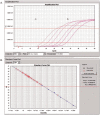
Detecting plasma Epstein-Barr virus DNA to diagnose postradiation nasopharyngeal skull base lesions in nasopharyngeal carcinoma patients: a prospective study
- PMID: 22237037
- PMCID: PMC3777480
- DOI: 10.5732/cjc.011.10279
Detecting plasma Epstein-Barr virus DNA to diagnose postradiation nasopharyngeal skull base lesions in nasopharyngeal carcinoma patients: a prospective study
Abstract
The diagnosis of postradiation nasopharyngeal skull base lesions in petients with nasopharyngeal carcinoma (NPC) is still a tough problem in clinical practice. An early and accurate diagnosis is important for subsequent management. We prospectively evaluated the diagnostic value of plasma Epstein-Barr virus(EBV) DNA in detecting postradiation nasopharyngeal skull base lesions in NPC patients. From July 2006 to September 2010, 90 patients with postradiation NPC (34 women and 56 men; median age: 42 years) met the selection criteria and were recruited in this study. All postradiation nasopharyngeal skull base lesions were found in the latest magnetic resonance imaging (MRI) examinations before endoscopic surgery, and the nasopharyngeal cavity was normal under flexible nasopharyngoscopy. Plasma EBV DNA detection was performed within 2 weeks before endoscopic surgery. A total of 90 endoscopic operations were successfully performed without any postoperative complications. Recurrences confirmed by postoperative pathology were found in 30 patients. The specificity, positive and negative predictive values of plasma EBV DNA detection were better than those of MRI. In addition, combining plasma EBV DNA detection with MRI improved the specificity and positive predictive values of MRI. Plasma EBV DNA detection followed by MRI would help to diagnose recurrence whereas MRI was unable. These results indicate that plasma EBV DNA is an effective and feasible biomarker for detecting postradiation nasopharyngeal skull base lesions in NPC patients.
Figures


Similar articles
-
Plasma Epstein-Barr virus DNA screening followed by ¹⁸F-fluoro-2-deoxy-D-glucose positron emission tomography in detecting posttreatment failures of nasopharyngeal carcinoma.Cancer. 2011 Oct 1;117(19):4452-9. doi: 10.1002/cncr.26069. Epub 2011 Mar 22. Cancer. 2011. PMID: 21437892
-
Noninvasive diagnosis of nasopharyngeal carcinoma: nasopharyngeal brushings reveal high Epstein-Barr virus DNA load and carcinoma-specific viral BARF1 mRNA.Int J Cancer. 2006 Aug 1;119(3):608-14. doi: 10.1002/ijc.21914. Int J Cancer. 2006. PMID: 16572427
-
Prognostic value of plasma Epstein-Barr virus DNA level during posttreatment follow-up in the patients with nasopharyngeal carcinoma having undergone intensity-modulated radiotherapy.Chin J Cancer. 2017 Nov 7;36(1):87. doi: 10.1186/s40880-017-0256-x. Chin J Cancer. 2017. PMID: 29116021 Free PMC article.
-
Application of circulating plasma/serum EBV DNA in the clinical management of nasopharyngeal carcinoma.Oral Oncol. 2014 Jun;50(6):527-38. doi: 10.1016/j.oraloncology.2013.12.011. Epub 2014 Jan 15. Oral Oncol. 2014. PMID: 24440146 Review.
-
Clinical utility of circulating Epstein-Barr virus DNA analysis for the management of nasopharyngeal carcinoma.Chin Clin Oncol. 2016 Apr;5(2):18. doi: 10.21037/cco.2016.03.07. Chin Clin Oncol. 2016. PMID: 27121878 Review.
Cited by
-
Protein tyrosine kinase 6 is associated with nasopharyngeal carcinoma poor prognosis and metastasis.J Transl Med. 2013 Jun 9;11:140. doi: 10.1186/1479-5876-11-140. J Transl Med. 2013. PMID: 23758975 Free PMC article.
-
Design of InnoPrimers-Duplex Real-Time PCR for Detection and Treatment Response Prediction of EBV-Associated Nasopharyngeal Carcinoma Circulating Genetic Biomarker.Diagnostics (Basel). 2021 Sep 25;11(10):1761. doi: 10.3390/diagnostics11101761. Diagnostics (Basel). 2021. PMID: 34679459 Free PMC article.
-
As an independent unfavorable prognostic factor, IL-8 promotes metastasis of nasopharyngeal carcinoma through induction of epithelial-mesenchymal transition and activation of AKT signaling.Carcinogenesis. 2012 Jul;33(7):1302-9. doi: 10.1093/carcin/bgs181. Epub 2012 May 18. Carcinogenesis. 2012. PMID: 22610073 Free PMC article.
-
Development and Validation of a Prognostic Nomogram Based on Residual Tumor in Patients With Nondisseminated Nasopharyngeal Carcinoma.Technol Cancer Res Treat. 2020 Jan-Dec;19:1533033820957035. doi: 10.1177/1533033820957035. Technol Cancer Res Treat. 2020. PMID: 32945239 Free PMC article.
-
Potential Interest in Circulating miR-BART17-5p As a Post-Treatment Biomarker for Prediction of Recurrence in Epstein-Barr Virus-Related Nasopharyngeal Carcinoma.PLoS One. 2016 Sep 29;11(9):e0163609. doi: 10.1371/journal.pone.0163609. eCollection 2016. PLoS One. 2016. PMID: 27684719 Free PMC article.
References
-
- Shin HR, Masuyer E, Ferlay J, et al. Cancer in Asia—Incidence rates based on data in cancer incidence in five continents IX (1998–2002) Asian Pac J Cancer Prev. 2010;11:11–16. - PubMed
-
- Smee RI, Meagher NS, Broadley K, et al. Recurrent nasopharyngeal carcinoma: current management approaches. Am J Clin Oncol. 2010;33:469–473. - PubMed
-
- Ng SH, Chang JT, Ko SF, et al. MRI in recurrent nasopharyngeal carcinoma. Neuroradiology. 1999;41:855–862. - PubMed
-
- Liu T, Xu W, Yan WL, et al. FDG-PET, CT, MRI for diagnosis of local residual or recurrent nasopharyngeal carcinoma, which one is the best? A systematic review. Radiother Oncol. 2007;85:327–335. - PubMed
-
- Lu TX, Mai WY, Teh BS, et al. Initial experience using intensity-modulated radiotherapy for recurrent nasopharyngeal carcinoma. Int J Radiat Oncol Biol Phys. 2004;58:682–687. - PubMed
MeSH terms
Substances
LinkOut - more resources
Full Text Sources
Miscellaneous
