[Correlation between podoplanin-positive lymphatic microvessel density and CT characteristics of non-small cell lung cancer]
- PMID: 22237122
- PMCID: PMC5999964
- DOI: 10.3779/j.issn.1009-3419.2012.01.07
[Correlation between podoplanin-positive lymphatic microvessel density and CT characteristics of non-small cell lung cancer]
Abstract
Background and objective: It has been proven that ymphatic microvessel density (LMVD) was closely correlated with the lymphatic metastasis of non-small cell lung cancer (NSCLC). The aim of the present study is to explore the relationship between podoplanin-LMVD and multi-slice spiral computed tomography (MSCT) characteristics of NSCLC.
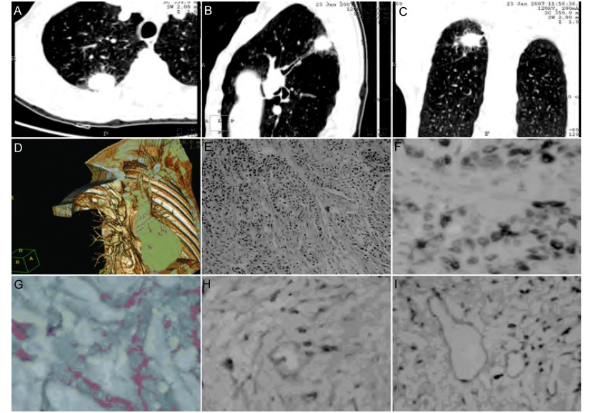
Methods: MSCT scanning was performed on 34 cases of NSCLC (squamous carcinoma, 15 cases; adenocarcinoma, 15 cases; and adenosquamous carcinoma, 4 cases) prior to operation. Clinical pathology results, including lymph node metastasis, were obtained. CT characteristics, such as shape of the edge, internal structure, and adjacent structures, were described. LMVD in the central and peripheral areas examined respectively using SP immunohistochemical technique were analyzed.
Results: Lymph node metastasis was found to be associated with LMVD in the peripheral areas. LMVD in the peripheral areas of the resected lesions, the MSCT findings of which included spinous process, pleural indentation, and carcinomatous lymphangitis, was higher than that of the lesions without these MSCT characteristics (P<0.05).
Conclusion: MSCT findings of spinous process, pleural indentation, or carcinomatous lymphangitis of NSCLC may suggest a higher level of tumor lymphangiogenesis with a higher risk of lymph node metastasis.
背景与目的: 现有的研究表明:肺癌微淋巴管密度(lymphatic microvessel density, LMVD)与淋巴结转移密切相关,但与肺癌多层螺旋CT(multi-slice spiral computed tomography, MSCT)的影像学改变的相关性尚不十分清楚。本研究通过podoplanin标记非小细胞肺癌患者手术标本微淋巴管并计数LMVD,观察患者肺癌病灶MSCT表现。
方法: 对34例非小细胞肺癌术前行MSCT检查,收集相关临床病理结果;评价MSCT表现(包括边缘形态、内部结构、邻近结构的CT征象);免疫组织化学SP法检测肿瘤组织中心区、周边区的LMVD。
结果: MSCT表现有棘状突起、胸膜凹陷征和癌性淋巴管炎的患者,其肺癌切除标本周围区LMVD均高于无上述表现者(P均 < 0.05)。
结论: MSCT出现棘状突起、胸膜凹陷征或癌性淋巴管炎表现提示更高的肿瘤淋巴管生成水平,具有更高的淋巴结转移风险。
Figures
Similar articles
-
Prognostic significance of VEGF-C expression in correlation with COX-2, lymphatic microvessel density, and clinicopathologic characteristics in human non-small cell lung cancer.Acta Biochim Biophys Sin (Shanghai). 2009 Mar;41(3):217-22. doi: 10.1093/abbs/gmp004. Acta Biochim Biophys Sin (Shanghai). 2009. PMID: 19280060
-
Detection of lymphangiogenesis in non-small cell lung cancer and its prognostic value.J Exp Clin Cancer Res. 2009 Feb 16;28(1):21. doi: 10.1186/1756-9966-28-21. J Exp Clin Cancer Res. 2009. PMID: 19216806 Free PMC article.
-
Correlation study between flash dual source CT perfusion imaging and regional lymph node metastasis of non-small cell lung cancer.BMC Cancer. 2020 Jun 12;20(1):547. doi: 10.1186/s12885-020-07032-8. BMC Cancer. 2020. PMID: 32532248 Free PMC article.
-
A meta-analysis of the relationship between lymphatic microvessel density and clinicopathological parameters in breast cancer.Bull Cancer. 2013 Mar;100(3):1-10. doi: 10.1684/bdc.2013.1719. Bull Cancer. 2013. PMID: 23501839 Review.
-
Lymphatic microvessel density as a prognostic factor in non-small cell lung carcinoma: a meta-analysis of the literature.Mol Biol Rep. 2012 May;39(5):5331-8. doi: 10.1007/s11033-011-1332-y. Epub 2011 Dec 14. Mol Biol Rep. 2012. PMID: 22167333
References
-
- Adachi Y, Nakamura H, Kitamura Y, et al. Lymphatic vessel density in pulmonary adenocarcinoma immunohistochemically evaluated with anti-podoplanin or anti-D2-40 antibody is correlated with lymphatic invasion or lymph node metastases. http://europepmc.org/abstract/med/17316411. Pathol Int. 2007;57(4):171–177. - PubMed
-
- Zhang GZ. Advances of imaging diagnostics of lung cancer. http://www.cqvip.com/Main/Detail.aspx?id=27681450 Chin J Lung Cancer. 2008;11(1):17–20. - PubMed
- 张 国桢. 肺癌的影像诊断学研究进展. http://www.cqvip.com/Main/Detail.aspx?id=27681450 中国肺癌杂志. 2008;11(1):17–20.
-
- Erasmus JJ, McAdams HP, Connolly JE. Solitary pulmonary nodules: Part Ⅱ. Evaluation of the indeterminate nodule. http://europepmc.org/abstract/MED/10682771 Radio graphics. 2000;20(1):59–66. - PubMed
-
- Weidner N. Tumor angiogenesis review of current applications in tumor prognostication. http://europepmc.org/abstract/med/7511250. Semin Diagn Pathol. 1993;10(4):302–313. - PubMed
Publication types
MeSH terms
Substances
LinkOut - more resources
Full Text Sources
Medical