Review
. 2012 Jan 12;3(1):e248.
doi: 10.1038/cddis.2011.123.

Cancer metabolism: current perspectives and future directions

Affiliations
Review

Cancer metabolism: current perspectives and future directions

C Muñoz-Pinedo et al. Cell Death Dis. .

Abstract

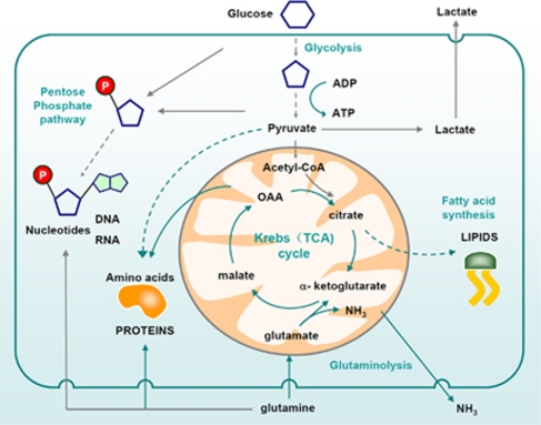
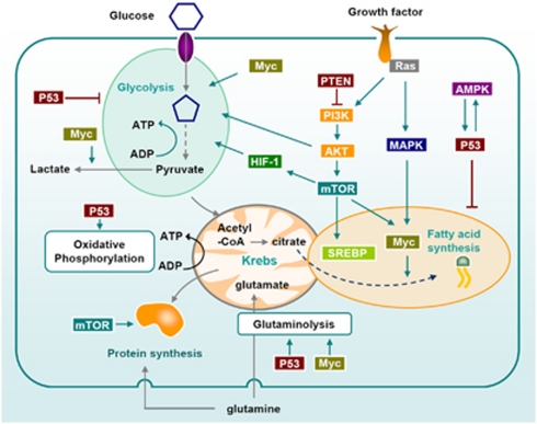
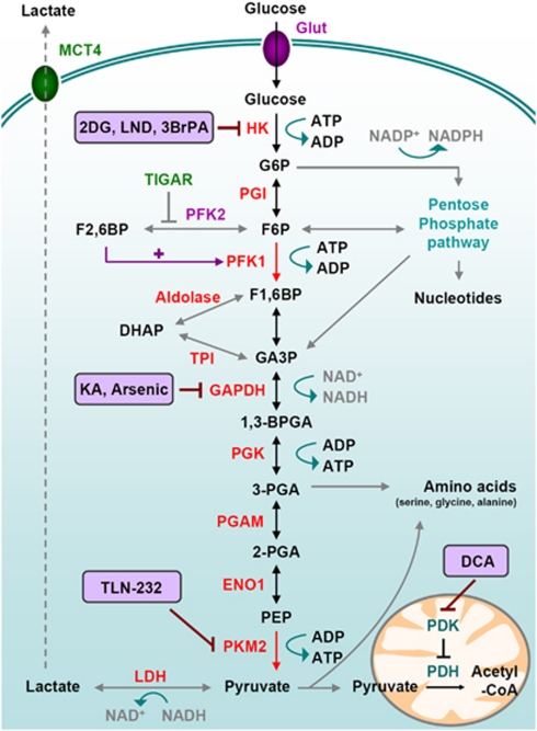
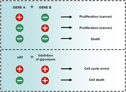
Cellular metabolism influences life and death decisions. An emerging theme in cancer biology is that metabolic regulation is intricately linked to cancer progression. In part, this is due to the fact that proliferation is tightly regulated by availability of nutrients. Mitogenic signals promote nutrient uptake and synthesis of DNA, RNA, proteins and lipids. Therefore, it seems straight-forward that oncogenes, that often promote proliferation, also promote metabolic changes. In this review we summarize our current understanding of how 'metabolic transformation' is linked to oncogenic transformation, and why inhibition of metabolism may prove a cancer's 'Achilles' heel'. On one hand, mutation of metabolic enzymes and metabolic stress sensors confers synthetic lethality with inhibitors of metabolism. On the other hand, hyperactivation of oncogenic pathways makes tumors more susceptible to metabolic inhibition. Conversely, an adequate nutrient supply and active metabolism regulates Bcl-2 family proteins and inhibits susceptibility to apoptosis. Here, we provide an overview of the metabolic pathways that represent anti-cancer targets and the cell death pathways engaged by metabolic inhibitors. Additionally, we will detail the similarities between metabolism of cancer cells and metabolism of proliferating cells.


Figures

Figure 1
Figure 1
Metabolism of proliferating cells. Proliferating cells require glucose which is converted to pyruvate through glycolysis. Pyruvate is converted to acetyl-CoA which enters the Krebs (TCA) cycle in the form of citrate. Alternatively, citrate is exported back to the cytosol to be used for lipid synthesis. Glucose can be also used as a source of carbon to produce ribose through the pentose phosphate pathway. Ribose-5-phosphate is then used to make RNA and DNA. Moreover, glycolytic intermediates such as pyruvate are used to produce non-essential amino acids such as alanine. Cells also require amino acids such as glutamine to make other amino acids and proteins. ‘Waste' is secreted in the form of lactate (mostly from glycolysis) and ammonia (from catabolism to amino acids)
Figure 2
Figure 2
Signaling pathways that regulate metabolism of proliferating and cancer cells. Growth factors influence metabolism through Ras and PI3K. Both PI3K/Akt and MAPK increase glycolysis. They also induce the upregulation of the transcription factor SREBP which promotes lipogenesis. mTOR, downstream of PI3K/Akt also plays a central role in the metabolic switch observed in highly proliferating cells: it activates protein translation, glycolysis (through HIF-1 dependent and independent pathways) and lipogenesis through the transcription factors SREBP and Myc. Myc is also the main oncogene implicated in glutamine addiction of cancer cells, through the upregulation of glutamate synthesis. It also contributes to the Warburg effect by increasing glycolysis and lactate production. AMPK activation, which is often impaired in tumors, allows the cells to switch their metabolism to catabolism when the nutrients are scarce. p53 regulates metabolism at multiple steps, notably through the upregulation of glutamate synthesis and inhibition of fatty acid synthesis and glycolysis,
Figure 3
Figure 3
Glucose metabolism in cancer cells. Glycolysis is a series of metabolic processes, driven by nine specific enzymes, by which one mole of glucose is catabolized to two moles of pyruvate, two moles of NADH with a net gain of two ATP. As indicated, several intermediates can fuel the Pentose Phosphate Pathway or lead to amino acid production. Accumulation of those intermediates is favored by the rate-limiting activity of PKM2. In cancer cells, pyruvate is further converted into lactate, thereby generating NAD+ from NADH. Pyruvate can be imported in the mitochondrial matrix to feed the TCA cycle. This step is controlled by Pyruvate Dehydrogenase Kinase (PDK) which can inactivate Pyruvate Dehydrogenase (PDH), therefore limiting the pyruvate conversion into acetyl-CoA and the further feeding of the TCA cycle. • Transporters: Glut: Glucose transporter; MCT: monocarboxylate transporter. • Glycolytic intermediates: G6P: Glucose-6-phosphate, F6P: fructose-6-phosphate; F1,6BP: fructose-1,6-bisphosphate; F2,6BP: fructose-2,6-bisphosphate; DHAP: dihydroxyacetone phosphate; GA3P: Glyceraldehyde-3-phosphate; 1,3-BPG: 1,3-bisphosphoglycerate; 3-PGA: 3-phosphoglycerate; 2-PG: 2-phosphoglycerate; PEP: phosphoenolpyruvate; • Enzymes: HK: hexokinase; PGI: phosphoglucoisomerase; PFK: Phosphofructokinase; TPI: triose phosphate isomerase; GAPDH: Glyceraldehyde 3-phosphate dehydrogenase; PGK: phosphoglycerate kinase; PGAM: phosphoglycerate mutase; ENO1: enolase 1; PK: pyruvate kinase; LDH: lactate dehydrogenase. • Chemical inhibitors are indicated in bold; 2DG: 2-Deoxy-Glucose; LND: Lonidamine; 3BrPA: 3-Bromopyruvate, KA: Koningic Acid; TLN-232 is a synthetic cyclic heptapeptide which targets PK; DCA: Dichloroacetate
Figure 4
Figure 4
Metabolic synthetic lethality. Synthetic lethality in organisms occurs when the simultaneous mutation of two genes is lethal, while mutation in each individual gene is not. Mutations in certain genes that occur frequently in cancer (for instance, p53) promote sensitivity to inhibition of specific metabolic pathways, which can be exploited to selectively target tumors with those mutations
Figure 5
Figure 5
Regulation of cell death by glucose metabolism. Nutrient availability regulates cell death induced by death receptors and by stimuli that kill through the mitochondrial pathway by regulating the antiapoptotic Bcl-2 family member Mcl-1, pro-apoptotic BH3-only proteins (Puma, Bim, Noxa and Bad) and c-FLIP. Glucose deprivation induces necrosis (not shown), caspase-8 mediated –but death receptor independent- apoptosis or mitochondrial apoptosis mediated by Noxa, Puma, Bad or Bim

References

    1. Warburg O. On respiratory impairment in cancer cells. Science. 1956;124:269. - PubMed
    1. Jones RG, Thompson CB. Tumor suppressors and cell metabolism: a recipe for cancer growth. Genes Dev. 2009;23:537–548. - PMC - PubMed
    1. DeBerardinis RJ, Sayed N, Ditsworth D, Thompson CB. Brick by brick: metabolism and tumor cell growth. Curr Opin Genetics Dev. 2008;18:54. - PMC - PubMed
    1. Michalek RD, Rathmell JC. The metabolic life and times of a T-cell. Immunological Reviews. 2010;236:190. - PMC - PubMed
    1. Fritz V, Fajas L. Metabolism and proliferation share common regulatory pathways in cancer cells. Oncogene. 2010;29:4369–4377. - PMC - PubMed

Publication types

MeSH terms