. 2012 Jan 12;3(1):e250.
doi: 10.1038/cddis.2011.133.

Prevention of neonatal oxygen-induced brain damage by reduction of intrinsic apoptosis

Affiliations

Prevention of neonatal oxygen-induced brain damage by reduction of intrinsic apoptosis

M Sifringer et al. Cell Death Dis. .

Abstract

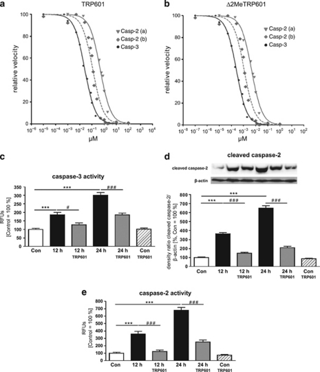
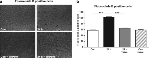
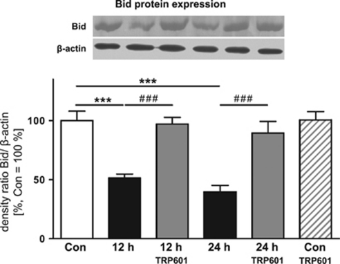
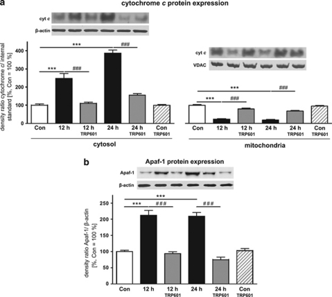
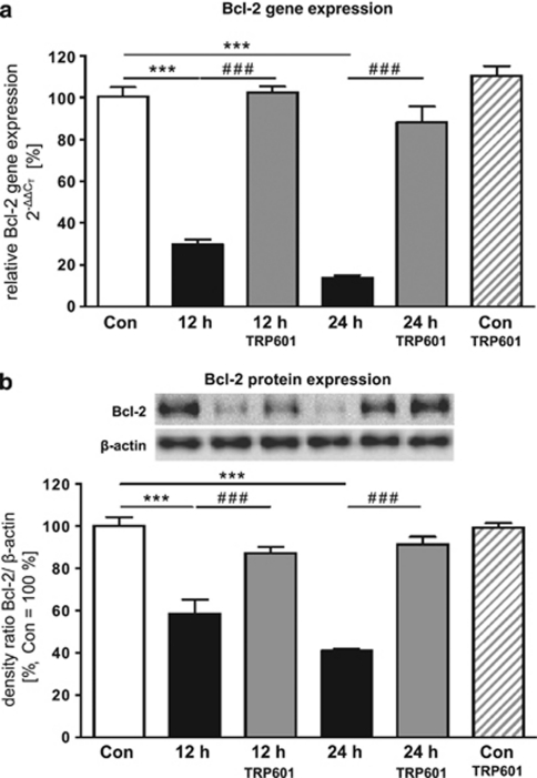
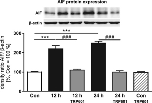
Within the last decade, it became clear that oxygen contributes to the pathogenesis of neonatal brain damage, leading to neurocognitive impairment of prematurely born infants in later life. Recently, we have identified a critical role for receptor-mediated neuronal apoptosis in the immature rodent brain. However, the contribution of the intrinsic apoptotic pathway accompanied by activation of caspase-2 under hyperoxic conditions in the neonatal brain still remains elusive. Inhibition of caspases appears a promising strategy for neuroprotection. In order to assess the influence of specific caspases on the developing brain, we applied a recently developed pentapeptide-based group II caspase inhibitor (5-(2,6-difluoro-phenoxy)-3(R,S)-(2(S)-(2(S)-(3-methoxycarbonyl-2(S)-(3-methyl-2(S)-((quinoline-2-carbonyl)-amino)-butyrylamino)propionylamino)3-methylbutyrylamino)propionylamino)-4-oxo-pentanoic acid methyl ester; TRP601). Here, we report that elevated oxygen (hyperoxia) triggers a marked increase in active caspase-2 expression, resulting in an initiation of the intrinsic apoptotic pathway with upregulation of key proteins, namely, cytochrome c, apoptosis protease-activating factor-1, and the caspase-independent protein apoptosis-inducing factor, whereas BH3-interacting domain death agonist and the anti-apoptotic protein B-cell lymphoma-2 are downregulated. These results coincide with an upregulation of caspase-3 activity and marked neurodegeneration. However, single treatment with TRP601 at the beginning of hyperoxia reversed the detrimental effects in this model. Hyperoxia-mediated neurodegeneration is supported by intrinsic apoptosis, suggesting that the development of highly selective caspase inhibitors will represent a potential useful therapeutic strategy in prematurely born infants.


Figures

Figure 1
Figure 1
Dose–response plots for inhibition of caspase-2 and -3 with TRP601 and its active metabolite. TRP601 (a) and Δ2Me-TRP601 (b) were added to recombinant caspase-2 (gray curves) and caspase-3 (black curves) to determine initial enzyme velocity and IC50 values in chromogenic microplate assay. TRP601 and Δ2Me-TRP601 were added simultaneously to substrates (plain curves; IC50/TRP601/Casp3=25.58±3.1 nM; IC50/Δ2Me-TRP601/Casp3=0.39±0.11 nM; IC50/TRP601/Casp2(a)=471.8±91.3 nM; IC50/Δ2Me-TRP601/Casp2(a)=7.4±3.18 nM) or alternatively 45 min before substrates (dotted curves; IC50/TRP601/Casp2(b)=115.2±39.12 nM; IC50/Δ2Me-TRP601/Casp2(b)=2.67±1.46 nM). (c) Six-day-old Wistar rat pups were either injected with the caspase inhibitor TRP601 (1 mg/kg bodyweight, i.p.) or vehicle control before exposure to 80% O2. After 12 or 24 h, animals were killed, transcardially perfused with PBS, and brain samples were collected in order to perform a fluorometric caspase-3 activity assay. Measurements of hydrolysis of Ac-DEVD-AMC at 460 nm resulted in a highly significant upregulation of caspase-3 activity under hyperoxic conditions, whereas single treatment with TRP601 significantly decreased caspase-3 activity to control levels after 12 and 24 h. Bars represent mean±S.E.M. (thalamus, n=6/group, normalized to control animals (21% O2) ***P<0.001, #P<0.05, ###P<0.001, two-way ANOVA). (d) Inhibition of caspase-2 by TRP601 in vivo (1 mg/kg bodyweight i.p.) lead to decreased protein expression of caspase-2 in thalamus from treated animals, whereas under normoxic conditions TRP601 had no effect. Data are normalized to levels of rat pups exposed to normoxia (control=100%). Representative western blot images of caspase-2 and β-actin are shown for thalamus. Bars represent mean ±S.E.M. (n=8/group, ***P<0.001, ###P<0.001, one-way ANOVA). (e) Six-day-old Wistar rat pups were either injected with the caspase inhibitor TRP601 (1 mg/kg bodyweight, i.p.) or vehicle control before exposure to 80% O2. After 12 or 24 h, animals were killed, transcardially perfused with PBS, and brain samples were collected in order to perform a fluorometric caspase-2 activity assay. Measurements of hydrolysis of VDVAD-AFC at 505 nm resulted in a highly significant upregulation of caspase-2 activity under hyperoxic conditions, whereas single treatment with TRP601 significantly decreased caspase-2 activity to control levels after 12 and 24 h. Bars represent mean ±S.E.M. (thalamus, n=6/group, normalized to control animals (21% O2) ***P<0.001, ###P<0.001, two-way ANOVA)
Figure 2
Figure 2
TRP601 ameliorates neuronal cell death in vivo. (a) Representative photomicrographs (original magnification × 400) of Fluoro-Jade B-stained 10-μm sections from the thalamus of seven-day-old rats, which were treated without or with TRP601 and were kept under room air (CON) or hyperoxic (24 h) conditions for 24 h. (b) Quantification of 16 brain regions indicates a highly significant reduction of Fluoro-Jade B-positive degenerated neurons after TRP601 treatment. Bars represent mean ±S.E.M. (n=8–12/group, ***P<0.001, ###P<0.001, one-way ANOVA)
Figure 3
Figure 3
Elevated oxygen concentrations induce Bid activation. Western blot from thalamic proteins and subsequent densitometric analysis of full-length Bid demonstrates a significant downregulation of Bid after hyperoxia (12 or 24 h, black bars), which is reversed by TRP601 treatment (gray bars, 1 mg/kg, i.p.), TRP601 under room air conditions showed no significant regulation (dashed bar). The densitometric data represent the ratio of the density of the Bid band to the corresponding β-actin band. Data are normalized to levels of rat pups exposed to normoxia (control=100% bars represent mean±S.E.M., n=7–9/group, ***P<0.001,###P<0.001, one-way ANOVA). Blots are representative of a series of three blots
Figure 4
Figure 4
TRP601 treatment inhibits hyperoxia-induced intrinsic apoptosis. (a) Western blot analysis from thalamic cytosolic and mitochondrial protein fractions implies a shift of cytochrome c from mitochondria to the cytosol under hyperoxic conditions that could be prevented by a single TRP601 treatment (1 mg/kg, i.p.). (b) Western blot analysis of Apaf-1 expression in thalamic brain samples displays a significant upregulation after 12 and 24 h hyperoxia, which was blocked by TRP601. The densitometric data represent the density ratio of relevant individual bands to the corresponding internal standard band (β-actin or VDAC). Data are normalized to levels of rat pups exposed to normoxia (control 100% bars represent mean±S.E.M., n=6/group, ***P<0.001, ###P<0.001, one-way ANOVA compared with respective controls). Blots are representative of a series of three blots
Figure 5
Figure 5
TRP601 restores Bcl-2 expression under hyperoxic conditions. (a) Quantitative analysis of mRNA expression by real-time PCR showed a marked reduction of Bcl-2 mRNA expression in thalamic samples of rat pups that were kept for 12 and 24 h under hyperoxia (black bars), whereas TRP601 treatment restores Bcl-2 expression to control levels (gray bars). Application of TRP601 under room air (control, dashed bar) showed no significant regulation on Bcl-2 mRNA expression. (b) The analysis of Bcl-2 protein expression by western blot showed a similar expression pattern. The protein expression of Bcl-2 is significantly decreased after 12 and 24 h; the single application of TRP601 could restore the Bcl-2 protein expression almost up to control level. The densitometric data represent the ratio of the density of the Bcl-2 band to the corresponding β-actin band. Data are normalized to levels of rat pups exposed to normoxia (control 100% bars represent mean±S.E.M., thalamus, n=6–8/group, ***P<0.001, ###P<0.001, one-way ANOVA compared with respective controls). Blots are representative of a series of three blots
Figure 6
Figure 6
Caspase-independent hyperoxia-induced apoptosis is regulated by TRP601 treatment. Densitometric quantification of AIF expression by western blotting demonstrates a significant upregulation of AIF expression in thalamic samples of hyperoxia-treated rat pups after 12 and 24 h, which is prevented by TRP601 treatment. The densitometric data represent the ratio of the density of the AIF band to the corresponding β-actin band. Data are normalized to levels of rat pups exposed to normoxia (control 100% bars represent mean±S.E.M.; n=6/group, ***P<0.001, ###P<0.001, one-way ANOVA compared with respective controls). Blots are representative of a series of three blots

Similar articles

Cited by

References

    1. Johnson S, Wolke D, Hennessy E, Marlow N. Educational outcomes in extremely preterm children: neuropsychological correlates and predictors of attainment. Dev Neuropsychol. 2011;36:74–95. - PubMed
    1. Keller M, Felderhoff-Mueser U, Lagercrantz H, Dammann O, Marlow N, Hüppi P, et al. Policy benchmarking report on neonatal health and social policies in 13 European countries. Acta Paediatr. 2010;99:1624–1629. - PubMed
    1. Felderhoff-Mueser U, Sifringer M, Polley O, Dzietko M, Leineweber B, Mahler L, et al. Caspase-1-processed interleukins in hyperoxia-induced cell death in the developing brain. Ann Neurol. 2005;57:50–59. - PubMed
    1. Gerstner B, Sifringer M, Dzietko M, Schüller A, Lee J, Simons S, et al. Estradiol attenuates hyperoxia-induced cell death in the developing white matter. Ann Neurol. 2007;61:562–573. - PubMed
    1. Schmitz T, Ritter J, Mueller S, Felderhoff-Mueser U, Chew L-J, Gallo V. Cellular changes underlying hyperoxia-induced delay of white matter development. J Neurosci. 2011;31:4327–4344. - PMC - PubMed

Publication types

MeSH terms