. 2012 Feb 15;72(4):928-38.
doi: 10.1158/0008-5472.CAN-11-2863. Epub 2012 Jan 11.

Antigen-specific CD4(+) T cells regulate function of myeloid-derived suppressor cells in cancer via retrograde MHC class II signaling

Affiliations

Antigen-specific CD4(+) T cells regulate function of myeloid-derived suppressor cells in cancer via retrograde MHC class II signaling

Srinivas Nagaraj et al. Cancer Res. .

Abstract

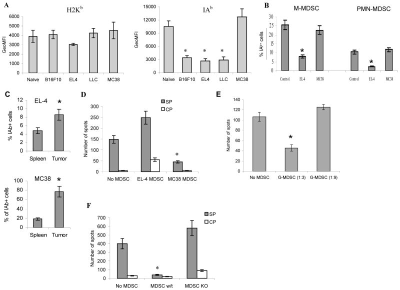
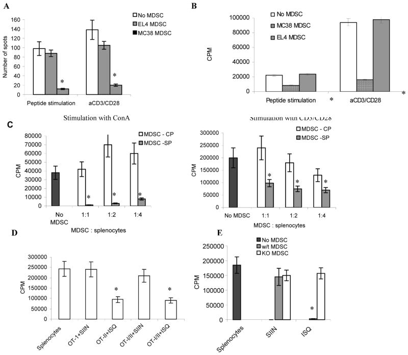
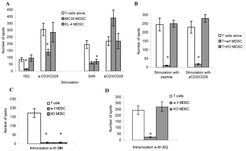
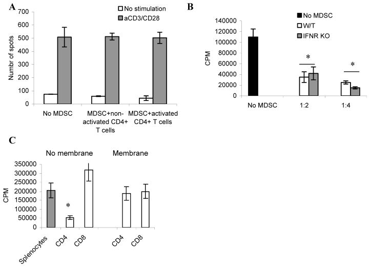
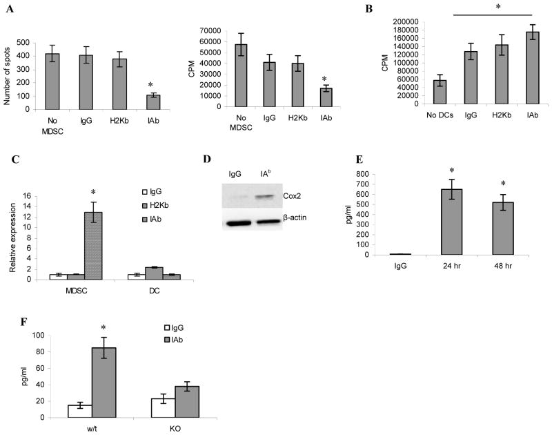
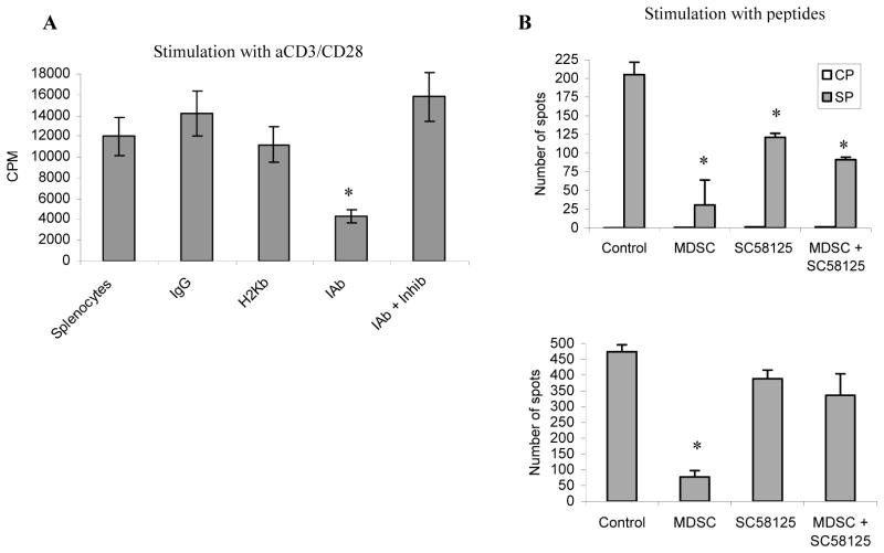
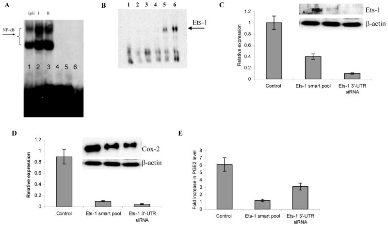
Myeloid-derived suppressor cells (MDSC) play a major role in cancer-related immune suppression, yet the nature of this suppression remains controversial. In this study, we evaluated the ability of MDSCs to elicit CD4(+) T-cell tolerance in different mouse tumor models. In contrast to CD8(+) T-cell tolerance, which could be induced by MDSCs in all the tumor models tested, CD4(+) T-cell tolerance could be elicited in only one of the models (MC38) in which a substantial level of MHC class II was expressed on MDSCs compared with control myeloid cells. Mechanistic investigations revealed that MDSCs deficient in MHC class II could induce tolerance to CD8(+) T cells but not to CD4(+) T cells. Unexpectedly, antigen-specific CD4(+) T cells (but not CD8(+) T cells) could dramatically enhance the immune suppressive activity of MDSCs by converting them into powerful nonspecific suppressor cells. This striking effect was mediated by direct cell-cell contact through cross-linking of MHC class II on MDSCs. We also implicated an Ets-1 transcription factor-regulated increase in expression of Cox-2 and prostaglandin E2 in MDSCs in mediating this effect. Together, our findings suggest that activated CD4(+) T cells that are antigen specific may enhance the immune suppressive activity of MDSCs, a mechanism that might serve normally as a negative feedback loop to control immune responses that becomes dysregulated in cancer.


Figures

Figure 1
Figure 1. MDSC-inducible CD4+ T-cell tolerance depends on MHC class II
A. Expression of IAb and H2Kb on MDSC. Expression of H2Kb (left panel) and IAb (right panel) was measured as geometric mean fluorescence intensity (MFI) within gated population of spleen Gr-1+CD11b+ cells. Mean and SD from at least 4 mice in each model are shown. * - statistically significant differences from control (p<0.05). B. The proportion of IAb+ cells among the populations of CD11b+Ly6C+Ly6G M-MDSC or CD11b+Ly6ClowLy6G+ PMN-MDSC from spleens of tumor-bearing or naïve mice. Each group includes four mice. C. The proportion of IAb+ cells among Gr-1+CD11b+ MDSC isolated from spleens and tumor tissues. To obtaine comparable results both spleens and tumors were treated with collagenase to collect cells. Each group includes four mice. D. OT-II splenocytes were cultured for 48 hr with MDSC from MC38 (MC38 MDSC) or EL-4 (EL-4 MDSC) TB mice at 3:1 ratio in the presence of specific (SP) or control (CP) peptides. The number of IFN-γ producing cells was calculated in triplicate in ELISPOT assay and presented as mean ± SD per 2×10⁵ cells. Each group included three mice. * - statistically significant differences from splenocytes incubated without MDSC (p<0.05). E. CD11b+Ly6G+Ly6Clo PMN-MDSC were sorted from spleens of MC38 TB mice and added to OT-II splenocytes at indicated ratios. Cells were incubated with control or specific peptide and ELISPOT assay was performed as described in Fig. 1D. Experiments were performed in triplicates. Numbers of spots in the presence of control peptide were <5 (not shown). * statistically significant differences from values without MDSC (p<0.05). F. MDSC isolated from C57/BL6 wild-type (w/t) and IAb knockout (KO) MC38 TB mice were cultured with OT II splenocytes and IFN-γ production was measured in ELISPOT assay as described in Fig. 1D.
Figure 2
Figure 2. CD4+ T cells cause conversion of MDSC to non-specific suppressors
A,B. CD4+ T-cell tolerance in vivo. OT-II CD4+ T cells were transferred i.v. into naïve C57BL/6 mice (5×10⁶ cells per mouse). Two days later mice were injected i.v. with 5×10⁶ MDSC from MC38 or EL-4 TB mice. On the same day, mice were immunized with 100 μg ISQ peptide in 100μl IFA. Ten days later, lymph node cells were isolated, stimulated with control or specific peptides or CD3/CD28 antibodies. IFN-γ production was evaluated in ELISPOT assay (A) and cell proliferation by 3H-thymidine uptake (B). The values in cells stimulated with control peptides were subtracted from values in cells stimulated with specific peptide. Each experiment was performed in triplicate and repeated twice. C. OT-II splenocytes and MDSC from MC38 tumor bearing mice were cultured together at a 3:1 ratio with control peptide (CP) or specific peptide (SP). 48 hr later, MDSC were isolated and added to naïve splenocytes stimulated with either ConA (5μg/ml) or CD3/CD28 (0.5μg/5μg/ml) antibody. Cell proliferation was assessed in triplicate in 3H-thymidine uptake. Two experiments with the same results were performed. D. OT-II CD4+ and OT-I CD8+ T cells were mixed at 1:1 ratio and cultured together with MDSC from MC38 tumor bearing mice at a 3:1 ratio. Specific peptides SIIN and ISQ were added as indicated. MDSC were isolated 48 hr later and cultured with naive splenocytes stimulated with CD3/CD28 antibody. Cell proliferation was evaluated as described in Fig. 2A. E. Experiments were performed exactly as described in Fig. 2D. MDSC were isolated from MC38 wild-type (w/t) and IAb knockout (KO) mice.
Figure 3
Figure 3. Conversion of MDSC to non-specific suppressors in vivo
A, B. 3×10⁶ OT-II CD4+ and 3×10⁶ OT-I CD8+ T cells were mixed together and injected i.v. into C57BL/6 recipient mice. Mice received MDSC from either EL-4 or MC38 tumor bearing mice two days later (A). Alternatively, MDSC from IAb knockout (KO) MC38 TB mice were used (B). The mice were immunized with ISQ or SIIN peptide as indicated. Eight-ten days later lymph node cells were isolated and stimulated with corresponding peptide or with anti-CD3/CD28 antibody. IFN-γ ELISPOT assay was performed in triplicate. Three experiments with the same results were performed. C, D. 3×10⁶ OT-II CD4+ and 3×10⁶ OT-I CD8+ T cells were mixed together and injected i.v. into C57BL/6 recipient mice. MDSC were isolated from wild-type (w/t) and IAb knockout (KO) MC38 TB mice and were transferred i.v. into recipient mice. These mice were immunized with either OT-I peptide-SIIN (C) or OT-II peptide ISQ (D). 8–10 days later lymph node cells were isolated and restimulated with corresponding peptides. IFN-γ ELISPOT assay was performed. Each experiment included three mice and each assay was performed in triplicate. Mean ± SD from all experiments are shown. * - statistically significant differences from values of T-cell activity without the presence of MDSC (p<0.01).
Figure 4
Figure 4. Mechanism of MDSC conversion to non-specific suppressors
A. Splenocytes from OT-II mice were cultured for 48 hr in complete medium (non-activated T cells) or in the presence 5 μg/ml ConA (activated T cells). T cells were then isolated and incubated for 48 hr with MDSC from MC38 TB mice in the absence of peptides. MDSC were then isolated and added to naïve splenocytes, stimulated with CD3/CD28 antibodies. IFN-γ production was assessed in ELISPOT assay. Experiment was performed in triplicate. Mean±SD in shown. B. MDSC were isolated from wild-type or IFN-γR knockout MC38 tumor bearing mice and cultured with OT-II CD4+ T cells in the presence of the specific peptide. MDSC were isolated 48 hr later and tested in their ability to suppress CD3/CD28 inducible activation of naïve T cells using different MDSC : splenocytes ratios. Proliferation was measured by 3H-thymidine uptake. Two experiments were performed. Each experiment was done in triplicates. * - statistically significant differences from values of T-cell proliferation without the presence of MDSC (p<0.05). C. OT-II and OT-I splenocytes were cultured with MDSC from MC38 mice at a 3:1 ratio in the presence of corresponding specific peptides. MDSC were either added to splenocytes directly (no membrane) or cultured on a semi-permeable (0.45 μm) membrane. MDSC were isolated 48 hr later and added to naive splenocytes at a 1:3 ratio. Cells were stimulated with CD3/CD28 specific antibody and proliferation was measured in 3H-thymidine uptake. Mean ± SD from three experiments (each experiment in triplicate) is shown. * - statistically significant differences from values of T-cell activity without the presence of MDSC (p<0.01).
Figure 5
Figure 5. Ligation of IAb on MDSC convert these cells to non-specific suppressors
A. MDSC isolated from MC38 TB mice were cultured for 48 hr with immobilized IgG, H2Kb, or IAb antibodies. MDSC were then harvested and added to naïve splenocytes stimulated with CD3/CD28 antibodies. IFN-γ ELISPOT (left panel) and proliferation (right panel) assays were performed. Cumulative results from 4 experiments (each in triplicate) are shown. * - statistically significant differences from values of T-cell activity without the presence of MDSC (p<0.05). B. Experiments were performed essentially as in Fig. 5A except DCs were used instead of MDSC. C. MDSC isolated from MC38 TB mice or bone marrow derived DCs were cultured with immobilized IgG, H2Kb, or IAb antibodies. 4hr later, RNA was isolated and relative expression of Cox-2 was analyzed using qRT PCR. Three experiments, in triplicate, were performed. D. MDSC from MC38 TB mice were cultured on plates with immobilized IgG or IAb antibody for 24 hr. Whole cell lysate was collected and Cox2 level was detected in Western blot. E. MDSC isolated from MC38 TB mice were cultured with immobilized IgG or IAb antibody for 24 or 48h. The concentration of PGE2 was measured in supernatants in ELISA. Two experiments (each in triplicate) with the same results were performed. The results of one experiment are shown. * - statistically significant differences from IgG control (p<0.001). F. Experiments were performed as described above. MDSC were isolated from wild-type and IAb KO mice and cultured for 24 hr with either immobilized control IgG or IAb antibody. PGE2 was measured in supernatants using ELISA. Two experiments, in triplicate, were performed. * - statistically significant differences from values in MDSC cultured with IgG (p<0.01).
Figure 6
Figure 6. CD4+ inducible conversion of MDSC is mediated by Cox2/PGE2
A. MDSC isolated from MC38 TB mice were incubated with immobilized IgG, H2Kb, or IAb antibodies for 48 hr with and without 5μM COX-2 inhibitor (SC58125) as indicated. After incubation MDSC were isolated, washed, and cultured with naive splenocytes stimulated with CD3/CD28 antibodies. Proliferation was measured by 3[H]-thymidine uptake in triplicate. Typical result of three experiments is shown. * - statistically significant differences from values of splenocytes cultured without MDSC (p<0.05). B. MDSC from MC38 TB mice were used in experimental model of CD4+ T-cell tolerance as described in Fig. 1D, E. SC5815 (10 mg/kg) was injected was injected i.p. on days 1, 3, 5, and 7 after MDSC administration. Lymph node cells were collected on day 10 and stimulated with control peptide (CP), specific peptide (SP) or CD3/CD28 antibodies. IFN-γ production was evaluated in ELISPOT assay. Experiment was performed in triplicates.
Figure 7
Figure 7. Up-regulation of Ets-1 in MDSC by IAb cross-linking on MDSC is responsible for increased production of PGE2
A. MDSC were isolated from spleens of MC38 TB mice and incubated for 16 hr with immobilized IgG, H2Kb, or IAb specific antibodies. NF-κB activity was measured using EMSA. Lines 1–3 NF-κB specific probe: 1 – MDSC incubated with IgG, 2 – H2Kb or 3 – IAb specific antibody; Lines 4–6 controls: 4 – NF-κB probe alone; 5 – MDSC after incubation with IAb antibody in the presence of 10-fold excess of unlabeled probe, 6 – the same in the presence 20-fold excess of unlabeled probe. B. MDSC were isolated from MC38 TB mice and cultured for 16 hr with immobilized I-Ab antibody or control IgG. Nuclear extracts were prepared and EMSA was performed using Ets-1 specific probe. Lanes: 1 - Ets-1 probe no cells; 2 - cells no probe; 3- cells cultured with IgG and labeled Ets-1 probe with 2x excess of unlabeled Ets-1 probe; 4 – as line 3 but without unlabeled probe; 5 – cells cultured with IAb and Ets-1 labeled probe with 2x excess of unlabeled Ets-1 probe; 6 – as line 5 but without unlabeled probe. Two experiments with the same result were performed. C. MDSC from MC38 TB mice were transfected with either control non-specific cyclophilin B pool siRNA, ETS1 specific 3′-UTR/ORF smart pool siRNA, or ETS1 specific 3′-UTR single siRNA. Cells were incubated with immobilized IAb for 48hr. RNA and proteins were isolated. Relative expression of ets-1 was analyzed (in triplicates) using qRT PCR (two experiments with the same results were performed). The level of Ets-1 protein was evaluated in Western blotting (inset). D. MDSC from MC38 TB mice were transfected with Ets-1 specific or scontrol siRNAs and incubated with immobilized IAb for 48h. Expression of cox2 mRNA (Mean ± st.d. of triplicates) and COX2 protein (inset) was measured by qRT-PCR and Western blot respectively. Two experiments with the same results were performed. E. PGE-2 levels were assessed in triplicate by ELISA. Mean ± st. dev. are shown. Three experiments with the same results were performed. In Fig. 7C–D differences between specific Ets-1 siRNA and control siRNA were statistically significant (p<0.05).

Similar articles

Cited by

References

    1. Gabrilovich D, Hurwitz A. Tumor induced immune suppression. New York: Springer; 2008.
    1. Gabrilovich DI, Nagaraj S. Myeloid-derived suppressor cells as regulators of the immune system. Nat Rev Immunol. 2009;9(3):162–74. - PMC - PubMed
    1. Ostrand-Rosenberg S, Sinha P. Myeloid-derived suppressor cells: linking inflammation and cancer. J Immunol. 2009;182(8):4499–506. - PMC - PubMed
    1. Sica A, Bronte V. Altered macrophage differentiation and immune dysfunction in tumor development. J Clin Invest. 2007;117(5):1155–66. - PMC - PubMed
    1. Solito S, Bronte V, Mandruzzato S. Antigen specificity of immune suppression by myeloid-derived suppressor cells. J Leukoc Biol. 2010 - PubMed

Publication types

Substances