Controlled Clinical Trial
. 2012 Jan 11;32(2):616-25.
doi: 10.1523/JNEUROSCI.2384-11.2012.

Tune it down to live it up? Rapid, nongenomic effects of cortisol on the human brain

Affiliations
Controlled Clinical Trial

Tune it down to live it up? Rapid, nongenomic effects of cortisol on the human brain

Florian Strelzyk et al. J Neurosci. .

Abstract

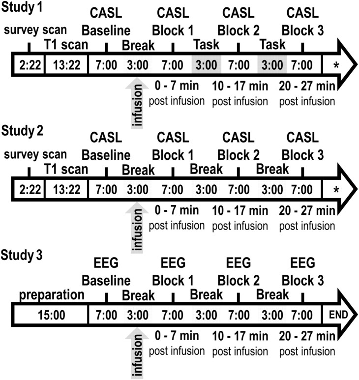
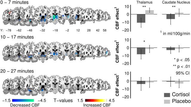
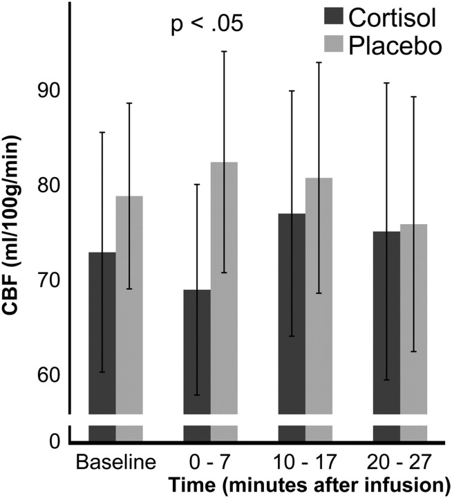
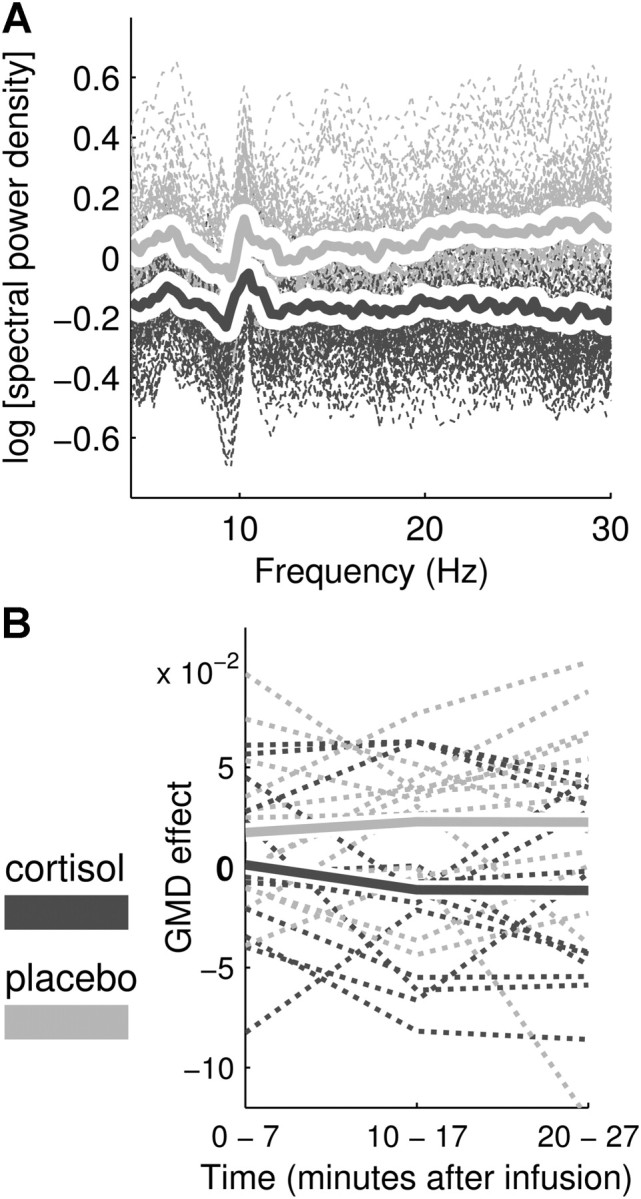
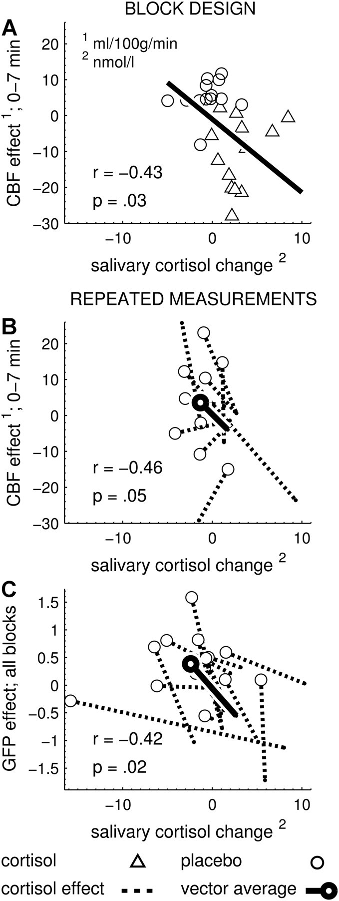
The stress hormone cortisol acts on the brain, supporting adaptation and time-adjusted coping processes. Whereas previous research has focused on slow emerging, genomic effects of cortisol, we addressed the rapid, nongenomic cortisol effects on in vivo neuronal activity in humans. Three independent placebo-controlled studies in healthy men were conducted. We observed changes in CNS activity within 15 min after intravenous administration of a physiological dose of 4 mg of cortisol (hydrocortisone). Two of the studies demonstrated a rapid bilateral thalamic perfusion decrement using continuous arterial spin labeling. The third study revealed rapid, cortisol-induced changes in global signal strength and map dissimilarity of the electroencephalogram. Our data demonstrate that a physiological concentration of cortisol profoundly affects the functioning and perfusion of the human brain in vivo via a rapid, nongenomic mechanism. The changes in neuronal functioning suggest that cortisol acts on the thalamic relay of background as well as on task-specific sensory information, allowing focus and facilitation of adaptation to challenges.


Figures

Figure 1.
Figure 1.
Sequence and duration (in minutes) of experimental procedures during data acquisition (Studies 1, 2, and 3). *The CASL measurements were succeeded by an M0 measurement (3 min, 25 s) and a T2-weighted anatomical measurement (2 min, 54 s) to further control for physiological abnormalities. The overall order and duration of the experimental measurement blocks (Baseline, Block 1, Block 2, and Block 3) were identical in all three studies.
Figure 2.
Figure 2.
Cortisol-induced rCBF changes (Study 1). Left, Statistical parametric maps for CBF changes for the interaction intervention × measurement (baseline corrected contrasts, cortisol vs placebo; p < 0.01, uncorrected) provide a descriptive overview of the spatial effect distribution and justify the interpretations of the ROI analyses. Locally distinct bilateral perfusion decrements in the thalamus and caudate nucleus (baseline corrected) after the intravenous infusion of cortisol (4 mg) for each measurement block (time, minutes after infusion). Right, Bilaterally averaged and baseline-corrected rCBF for significant ROI comparisons for all postinfusion measurement blocks [95% confidence interval CI)].
Figure 3.
Figure 3.
Cortisol-induced rCBF changes in the bilateral thalamus in Study 2. Cortisol induced a bilateral perfusion decrease in the thalamus during the first measurement block (0–7 min) after the infusion (95% confidence interval). This result replicates the thalamic perfusion decrease during the first measurement block of Study 1.
Figure 4.
Figure 4.
Cortisol-induced changes in global EEG markers (Study 3). A, ln-transformed and baseline-corrected FFT power spectra for all postinfusion measurement blocks, electrodes, and participants (dashed lines), with the average for cortisol (solid dark gray line) and placebo (solid light gray line) conditions. The cortisol-induced decrease in spectral power stretches across all frequency bins (4–30 Hz). B, Baseline-corrected GMD for all postinfusion measurement blocks (0–7 min, 10–17 min, 20–27 min) displayed for each participant (dashed lines) and averaged for cortisol (solid dark gray line) and placebo (solid light gray line) conditions.
Figure 5.
Figure 5.
Significant correlations between dependent variables and salivary cortisol changes. A, Study 1. Baseline-corrected thalamic blood flow of the first measurement block (ordinate) is negatively correlated with the change in salivary cortisol (abscissa, cortisol levels after MRI minus cortisol levels before MRI). This correlation is plotted across all participants, since cortisol and placebo groups contained different participants. B, Study 2. Baseline-corrected thalamic blood flow of the first measurement block is also negatively correlated with the change in salivary cortisol. This correlation is plotted as individual effect vectors because the cortisol and placebo conditions contained the same participants (repeated measurement study). C, Study 3. Baseline-corrected GFP values (ordinate) were averaged across the three postinfusion measurement blocks (because the effect of intervention was significant across all blocks) and showed a negative correlation with the salivary cortisol change (abscissa, cortisol levels after EEG minus cortisol levels before EEG).

Similar articles

Cited by

References

    1. Alsop DC, Detre JA. Reduced transit-time sensitivity in noninvasive magnetic resonance imaging of human cerebral blood flow. J Cereb Blood Flow Metab. 1996;16:1236–1249. - PubMed
    1. Bell AJ, Sejnowski TJ. An information-maximization approach to blind separation and blind deconvolution. Neural Comput. 1995;7:1129–1159. - PubMed
    1. Bloomfield P. Fourier analysis of time series: an introduction. New York: Wiley; 1976.
    1. Brunia CH. Neural aspects of anticipatory behavior. Acta Psychol (Amst) 1999;101:213–242. - PubMed
    1. Chatrian GE, Lettich E, Nelson PL. Modified nomenclature for the “10%” electrode system. J Clin Neurophysiol. 1988;5:183–186. - PubMed

Publication types

MeSH terms

LinkOut - more resources