Comparative Study
. 2012 Jan 11;32(2):626-38.
doi: 10.1523/JNEUROSCI.4254-11.2012.

Why do axons differ in caliber?

Affiliations
Comparative Study

Why do axons differ in caliber?

János A Perge et al. J Neurosci. .

Abstract

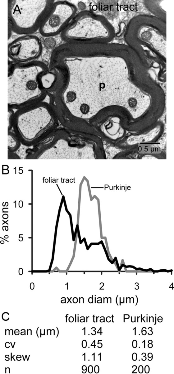
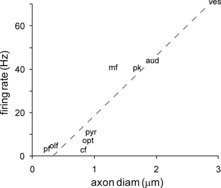
CNS axons differ in diameter (d) by nearly 100-fold (∼0.1-10 μm); therefore, they differ in cross-sectional area (d(2)) and volume by nearly 10,000-fold. If, as found for optic nerve, mitochondrial volume fraction is constant with axon diameter, energy capacity would rise with axon volume, also as d(2). We asked, given constraints on space and energy, what functional requirements set an axon's diameter? Surveying 16 fiber groups spanning nearly the full range of diameters in five species (guinea pig, rat, monkey, locust, octopus), we found the following: (1) thin axons are most numerous; (2) mean firing frequencies, estimated for nine of the identified axon classes, are low for thin fibers and high for thick ones, ranging from ∼1 to >100 Hz; (3) a tract's distribution of fiber diameters, whether narrow or broad, and whether symmetric or skewed, reflects heterogeneity of information rates conveyed by its individual fibers; and (4) mitochondrial volume/axon length rises ≥d(2). To explain the pressure toward thin diameters, we note an established law of diminishing returns: an axon, to double its information rate, must more than double its firing rate. Since diameter is apparently linear with firing rate, doubling information rate would more than quadruple an axon's volume and energy use. Thicker axons may be needed to encode features that cannot be efficiently decoded if their information is spread over several low-rate channels. Thus, information rate may be the main variable that sets axon caliber, with axons constrained to deliver information at the lowest acceptable rate.


Figures

Figure 1.
Figure 1.
Photoreceptor axon diameters vary with information capacity. A, Rod and cone axons in cross section (monkey fovea, courtesy of Y. Tsukamoto, Hyogo College of Medicine, Hyogo, Japan). Rod axons are numerous and thin, whereas cone axons are sparse and thick. Rod transmits a binary signal (0 or 1 photon) to modulate transmitter release at a single ribbon synapse. The axon caliber is fine and both narrowly and symmetrically distributed. Cone axon transmits a finely graded signal to modulate transmitter release at ∼20 ribbon synapses. The axon is thicker and the distribution is negatively skewed with a tail toward finer axons. B, Distribution of axon diameters. The distribution includes three cell types with peak sensitivities to different wave lengths (S, M, and L). S cones carry less information than M and L cones (Garrigan et al., 2010) and are sparse (∼5% of the total). If the S axons are finer, as predicted, they might explain the skewed distribution. C, Summary statistics. cv, Coefficient of variation; n, number of fibers measured.
Figure 2.
Figure 2.
Four types of unmyelinated spiking axons show skewed distributions of diameter. Mean diameters vary by fourfold, skew varies by twofold. A, Parallel fibers from cerebellum show the least skew, hypothetically because they all serve a similar function and fire at similar mean rates. Their spread is approximately twofold greater than for rod axons. This might reflect differences among parallel fibers in mean activity level due to adaptive synapses. B, Olfactory receptor axons are twofold thicker than parallel fibers, and shows 37% greater skew. Each olfactory receptor expresses one of ∼1000 G-proteins, which in natural environments might be activated with different frequencies and intensities. Thus differences in information capacity among axons might explain the large skew. Some olfactory axons are immature and these are the finest fibers. Omitting them from the distribution would shift the lower cutoff rightward but not affect the skew. Unmyelinated fornix axons show nearly the same distribution as the olfactory fibers; nothing is known about their function. C, Unmyelinated axon segments of ganglion cells within the retina show a broader distribution than the olfactory fibers and a strong skew. This distribution is known to match the distribution of firing rates (Perge et al., 2009). Electron micrographs: A from rat; B, D from guinea pig. E, Summary statistics.
Figure 3.
Figure 3.
Medium caliber myelinated tracts of different function and length allocate space similarly. A, Fornix (hippocampus to mammillary body, ∼ 5 mm) contains mostly myelinated axons, plus some unmyelinated axons whose diameter distribution is shown in Figure 2. B, Pyramidal tract (anterior cortex to lower medulla and spinal cord, ≥ 23 mm). C, Optic nerve (eye to lateral geniculate nucleus, ∼17 mm). D, All three myelinated tracts show similar structure: same lower bound on fiber diameter; similar distribution of diameters, despite their different conduction distances. Electron micrographs from guinea pig. E, Summary statistics.
Figure 4.
Figure 4.
Foliar tract in cerebellum contains mostly thick axons. A, Purkinje cell axons (p), identified by extra-dense and extra-thick myelin sheath are thickest, but medium and fine axons are also present. B, Distribution of fiber diameters in foliar tract. Thickest fibers are Purkinje axons. C, Coefficient of variation and skew are small for Purkinje axons, consistent with their homogeneous function. Electron micrograph from rat.
Figure 5.
Figure 5.
Auditory axons are thick and uniform. A, Auditory axons are thick and uniform. Electron micrograph, rat. Note the different scale bar compared with the previous figures. B, Distribution of fiber diameter from the auditory nerve exemplified in A and also fibers only from basal cochlea (guinea pig, replotted from Gleich and Wilson, 1993). Note that basal axons distribute their diameters more narrowly and more symmetrically. This distribution is expected from our hypothesis because these axons share a narrower range of coding frequencies than the nerve as a whole. These fibers are also thicker than the overall population (Friede, 1984). Since these fibers encode the highest signal frequencies, they probably carry higher information rates. Thus, their greater thickness is also expected on the hypothesis that higher mean information rates require thicker axons, as noted by Friede (1984). C, Summary statistics.
Figure 6.
Figure 6.
Vestibular fibers are very thick and broadly distributed. Distribution of fiber diameters. Mean is greatest of all tracts surveyed, with long tail to extremely thick axons. Replotted from Gacek and Rasmussen (1961). Summary statistics: broad distribution expected because vestibular nerve reports from three different end-organs (utricle, sacculus, and macula), each likely to signal with somewhat different mean rates.
Figure 7.
Figure 7.
Distributions of axon caliber in invertebrate species resemble that of mammal. A, Insect ventral nerve cord. Electron micrograph, locust. B, Distribution of axon diameters from locust. C, Distribution of axon diameters from octopus chromatophore lobe. Note that shape of distribution resembles that for the guinea pig optic nerve. D, Summary statistics. Replotted from Camm (1986).
Figure 8.
Figure 8.
Energy capacities of various tracts in this study. A, Mitochondrial concentrations for all fiber groups studied. Unmyelinated axons invariably express a higher volume fraction than myelinated axons, which is consistent with the greater costs that unmyelinated axons must pay for sodium pumping. The photoreceptor axons, which conduct passively, are nearly devoid of mitochondria. B, Mitochondrial concentrations are similar for three medium-caliber, myelinated tracts. C, Capillary area correlates with mitochondrial volume, suggesting that energy capacity correlates with energy use.
Figure 9.
Figure 9.
Estimated mean firing rate versus mean axon diameter for nine different fiber tracts. For nine myelinated tracts, finer fibers use lower mean firing rates. See Results for references regarding estimated firing rates. pf, Parallel fiber; olf, olfactory axon; cf, climbing fiber; opt, optic axons; pyr, pyramidal tract axons; mf, mossy fiber; pk, Purkinje axon; aud, auditory axons; ves, vestibular axons.

Similar articles

Cited by

References

    1. Ahn HS, Black JA, Zhao P, Tyrrell L, Waxman SG, Dib-Hajj SD. Nav1.7 is the predominant sodium channel in rodent olfactory sensory neurons. Mol Pain. 2011;7:32. - PMC - PubMed
    1. Angelaki DE, Cullen KE. Vestibular system: the many facets of a multimodal sense. Annu Rev Neurosci. 2008;31:125–150. - PubMed
    1. Armstrong DM, Drew T. Discharges of pyramidal tract and other motor cortical neurones during locomotion in the cat. J Physiol. 1984;346:471–495. - PMC - PubMed
    1. Attwell D, Laughlin SB. An energy budget for signaling in the grey matter of the brain. J Cereb Blood Flow Metab. 2001;21:1133–1145. - PubMed
    1. Baker MR, Edgley SA. Non-uniform olivocerebellar conduction time in the vermis of the rat cerebellum. J Physiol. 2006;570:501–506. - PMC - PubMed

Publication types

MeSH terms

LinkOut - more resources