. 2012 Mar;86(6):3211-8.
doi: 10.1128/JVI.06810-11. Epub 2012 Jan 11.

Varicella-zoster virus infects human embryonic stem cell-derived neurons and neurospheres but not pluripotent embryonic stem cells or early progenitors

Affiliations

Varicella-zoster virus infects human embryonic stem cell-derived neurons and neurospheres but not pluripotent embryonic stem cells or early progenitors

Anna Dukhovny et al. J Virol. 2012 Mar.

Abstract

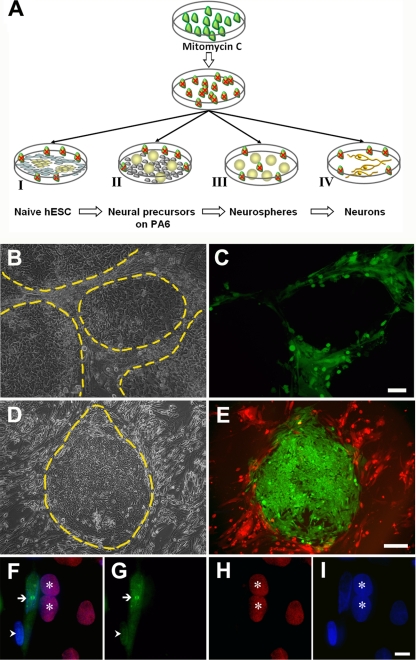
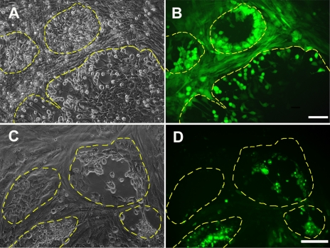
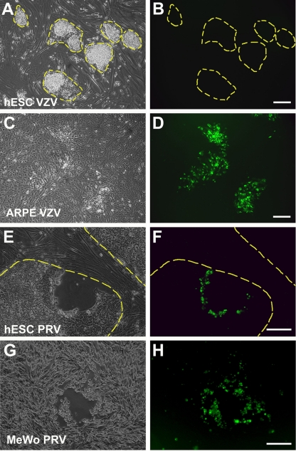
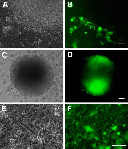
Pluripotent human stem cells are a powerful tool for the generation of differentiated cells that can be used for the study of human disease. We recently demonstrated that neurons derived from pluripotent human embryonic stem cells (hESC) can be infected by the highly host-restricted human alphaherpesvirus varicella-zoster virus (VZV), permitting the interaction of VZV with neurons to be readily evaluated in culture. In the present study, we examine whether pluripotent hESC and neural progenitors at intermediate stages of differentiation are permissive for VZV infection. We demonstrate here that VZV infection is blocked in naïve hESC. A block to VZV replication is also seen when a bacterial artificial chromosome (BAC) containing the VZV genome is transfected into hESC. In contrast, related alphaherpesviruses herpes simplex virus 1 (HSV-1) and pseudorabies virus (PrV) productively infect naïve hESC in a cell-free manner, and PrV replicates from a BAC transfected into hESC. Neurons differentiate from hESC via neural progenitor intermediates, as is the case in the embryo. The first in vitro stage at which permissiveness of hESC-derived neural precursors to VZV replication is observed is upon formation of "neurospheres," immediately after detachment from the inductive stromal feeder layer. These findings suggest that hESC may be useful in deciphering the yet enigmatic mechanisms of specificity of VZV infection and replication.


Figures

Fig 1
Fig 1
Naïve hESC are not infected by VZV. (A) Schematic of the infection of hESC and their neurally differentiated derivatives. VZV-infected MeWo or ARPE cells (green) were treated with mitomycin C to prevent proliferation and overrunning the hESC cultures (indicated by red x over cells). Mitotically inhibited VZV-MeWo cells were then mixed with one of four different targets: I, pluripotent hESC cells (yellow) growing on foreskin fibroblasts (gray); II, neurally induced hESC colonies (round yellow balls) on PA6 stromal feeders (gray); III, hESC-derived neurospheres (yellow balls) in suspension; or IV, hESC-derived neural precursors differentiated by plating on a laminin substrate (dark yellow neurons). (B and C) VZV-GFP-infected MeWo cells were added to hESC grown on human fibroblasts, and the live cultures were photographed 7 days later. The fibroblast feeders are infected with the virus and express GFP, whereas the hESC colonies are devoid of GFP expression. Panel B is a phase-contrast micrograph with several large hESC colonies demarcated by dashed yellow lines adjacent to infected, GFP-expressing feeder fibroblasts. Panel C is a combined fluorescence and phase-contrast micrograph. (D and E) hESC cells stably expressing GFP under the control of the EF1α promoter grown on a human fibroblast feeder layer were infected with mitomycin-treated VZV-RFP23-containing MeWo cells and imaged 3 days later. Again, the virus infected and labeled the feeders red, and the GFP-expressing hESC are not infected. Panel D is a phase-contrast micrograph of a large hESC colony demarcated by a dashed yellow line, and panel E shows only fluorescence. Red fluorescence is only observed outside the GFP-expressing hESC colonies. (F to I) Lack of evidence for VZV virion/capsid entry into undifferentiated hESC cells. Mitotically inhibited VZV-GFP23-containing ARPE cells were added to a culture of hESC. Two days later, cultures were fixed and immunostained for GFP (green) and Oct-4, a marker for pluripotent stem cells (red), and examined with a 100× oil-immersion objective. A strongly infected fibroblast displays GFP fluorescence in both its nucleus and cytoplasm (arrow), whereas an apparently more recently infected fibroblast (arrowhead) displays GFP only in its nucleus, and no ORF23-GFP is yet apparent in its cytoplasm. Pluripotent (Oct-4 immunopositive; red) hESC (asterisks) adjacent to the fibroblast whose cytoplasm is filled with GFP are completely devoid of GFP immunofluorescence. (F) Composite of all fluorescence channels; (G) GFP (ORF23) fluorescence only; (H) Oct-4 labeling only; (I) nuclear Hoechst staining (blue). Scale bars are 100 μm in panels B and C, 200 μm in panels D and E, and 20 μm in panels F to I.
Fig 2
Fig 2
Alphaherpesviruses HSV-1 and PrV productively infect naïve hESC. hESC grown on a feeder layer of mitotically inhibited human foreskin fibroblasts were infected with cell-free GFP-expressing HSV-1 (A and B) or PrV (C and D) and photographed 2 and 4 days after introduction of virus, respectively. Both the hESC (surrounded by dashed lines) and the surrounding feeder cells express GFP, indicating infection by the viruses. Many hESC show cytopathic effects after infection with these viruses, and cell-free plaques left by dead and detached hESC are seen in the middle of colonies infected with PrV. In panels A and C, micrographs are phase contrast only, whereas, in panels B and D, GFP fluorescence is shown. Scale bars, 100 μm.
Fig 3
Fig 3
VZV is unable to replicate in hESC when infection is bypassed by transfection of BAC DNA containing the VZV genome. hESC and ARPE cells were transfected with BACs containing VZV-GFP62 or PrV-GFP and photographed 6 days later. None of the hESC colonies displayed GFP, indicating VZV-GFP62 infection (A and B) in contrast to the many fluorescent foci resulting from viral replication in the ARPE cells (C and D). Transfection of PrV genome-containing BACs into hESC results in viral replication, as demonstrated by the GFP fluorescence after transfection with PrV-GFP (E and F). Transfection of PrV-GFP BACs in parallel to MeWo cells also leads to viral replication (G and H). Panels A, C, E, and G are phase-contrast micrographs, and panels B, D, F, and H are fluorescence photomicrographs of the same fields, with hESC colonies delineated by yellow dashed lines. Scale bars, 100 μm.
Fig 4
Fig 4
Neurospheres in suspension are the first stage of neural differentiation of hESC that support infection by VZV. (A and B) hESC cells were plated on PA6 stromal cells and differentiated for 14 days (shown diagrammatically in Fig. 1II). Mitomycin-treated VZV-GFP-infected MeWo cells were added to the coculture 10 to 12 days after hESC plating. Panels A and B show an hESC colony from a 14-day coculture with adjacent GFP-expressing MeWo cells. (The PA6 cells are murine and are not infected by VZV.) The neurally differentiating hESC colony is devoid of fluorescence. (C and D) hESC were cocultured with PA6 for 14 days as in panels A and B, and colonies were cut out and placed in suspension culture with VZV-GFP-infected MeWo cells (Fig. 1III). Three days postinfection, most of the neurospheres express GFP, indicating infection. (E and F) Neurospheres generated as described above were plated on laminin-coated coverslips and VZV-GFP-infected MeWo cells were added to the cultures immediately after adhesion of the neurosphere cells (Fig. 1IV). Three days after plating, the culture is a mixture of neural precursors and differentiated neurons bearing an extensive plexus of neurites. VZV infection of the neural cells is readily observed by GFP expression driven by an SV40 promoter. GFP diffusely fills the neurites and neuronal cell bodies. Panels A, C, and E are phase-contrast images, and panels B, D, and E are fluorescence images. Scale bars, 100 μm.

References

    1. Arvin AM, et al. 2010. Varicella-zoster virus T cell tropism and the pathogenesis of skin infection. Curr. Top. Microbiol. Immunol. 342:189–209 - PMC - PubMed
    1. Brokhman I, et al. 2009. Genetic modification of human embryonic stem cells with adenoviral vectors: differences of infectability between lines and correlation of infectability with expression of the coxsackie and adenovirus receptor. Stem Cells Dev. 18:447–456 - PubMed
    1. Butler JT, Hall LL, Smith KP, Lawrence JB. 2009. Changing nuclear landscape and unique PML structures during early epigenetic transitions of human embryonic stem cells. J. Cell. Biochem. 107:609–621 - PMC - PubMed
    1. Cowan CA, et al. 2004. Derivation of embryonic stem-cell lines from human blastocysts. N. Engl. J. Med. 350:1353–1356 - PubMed
    1. Decman V, Kinchington PR, Harvey SAK, Hendricks RL. 2005. Gamma interferon can block herpes simplex virus type 1 reactivation from latency, even in the presence of late gene expression. J. Virol. 79:10339–10347 - PMC - PubMed

Publication types