. 2012 Mar;86(6):3253-63.
doi: 10.1128/JVI.06648-11. Epub 2012 Jan 11.

The viral protein Tat can inhibit the establishment of HIV-1 latency

Affiliations

The viral protein Tat can inhibit the establishment of HIV-1 latency

Daniel A Donahue et al. J Virol. 2012 Mar.

Abstract

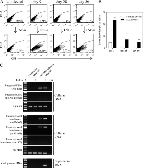
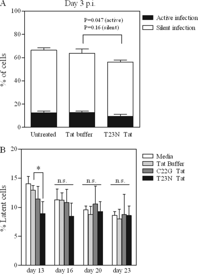
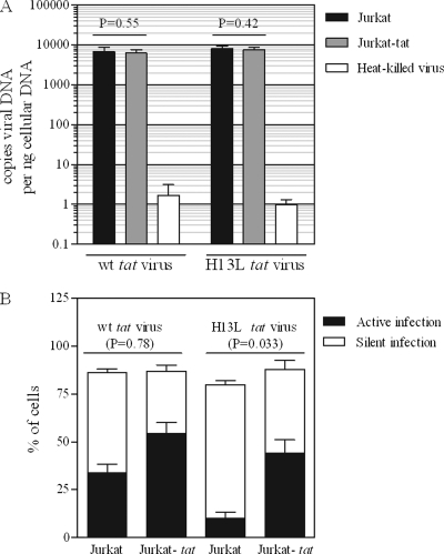
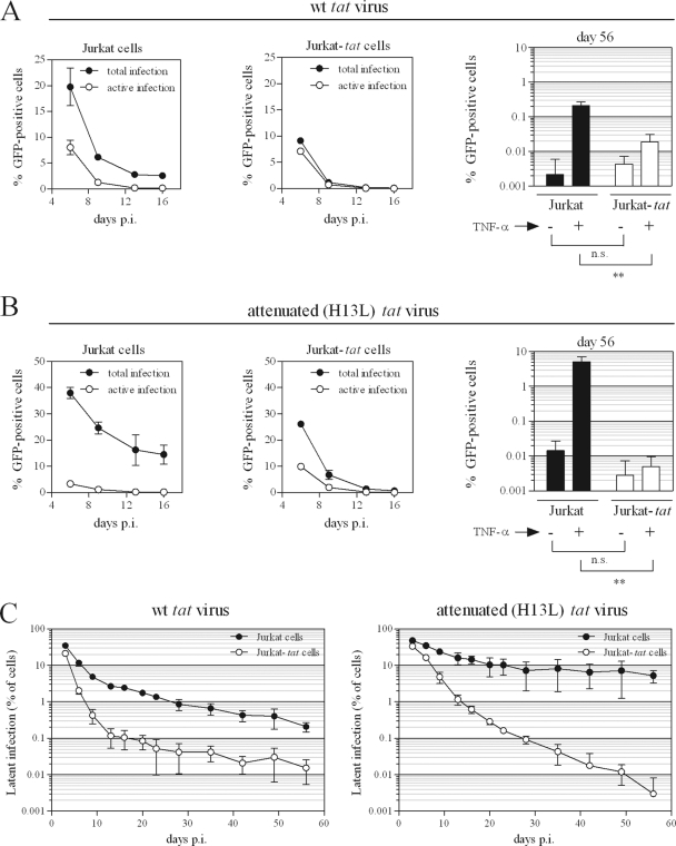
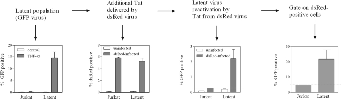
The establishment of HIV-1 latency can result from limiting levels of transcription initiation or elongation factors, restrictive chromatin modifications, transcriptional interference, and insufficient Tat activity. Since the viral protein Tat can counteract many of these factors, we hypothesized that the presence of exogenous Tat during infection might inhibit the establishment of latency. This was explored using a Jurkat model of latency establishment and reactivation. PCR and reverse transcriptase PCR (RT-PCR) confirmed the latent state in this model and showed evidence of transcriptional interference. To address our hypothesis, cells undergoing infection were first exposed to either purified recombinant Tat or a transactivation-negative mutant. Only the former resulted in a modest inhibition of the establishment of latency. Next, Jurkat cells stably expressing intracellular Tat were used in our latency model to avoid limitations of Tat delivery. Experiments confirmed that intracellular Tat expression did not affect the susceptibility of these cells to viral infection. Eight weeks after infection, Jurkat cells expressing Tat harbored up to 1,700-fold fewer (P < 0.01) latent viruses than Jurkat cells that did not express Tat. Additionally, Tat delivered by a second virus was sufficient to reactivate most of the latent population. Our results suggest that inhibition of the establishment of latent infection is theoretically possible. In a hypothetical scenario of therapy that induces viral gene expression during acute infection, activation of viruses which would otherwise have entered latency could occur while concurrent highly active antiretroviral therapy (HAART) would prevent further viral spread, potentially decreasing the size of the established latent reservoir.


Figures

Fig 1
Fig 1
Model of HIV-1 latency establishment and reactivation. Jurkat cells were infected with NL4-3-ΔE-EGFP or the attenuated tat (H13L) derivative and cultured for up to 8 weeks. Productively infected cells die by cytopathic effect, leaving only uninfected and latently infected cells, which can be reactivated with TNF-α and quantified by flow cytometry for viral EGFP. (A) Representative flow cytometry results for one of three independent experiments with attenuated tat virus. (B) Results of three independent experiments with attenuated or wild-type tat viruses; results represent mean ± standard deviation (SD). (C) PCR and RT-PCR characterization of uninfected or latently infected cells 28 days p.i. Integrated DNA was detected with Alu-gag primers; reactions with no Alu primer serve as a control to confirm that the Alu-gag band is derived from integrated DNA. Two different products of transcriptional interference were detected by RT-PCR, containing viral U3-PBS sequence that is derived from host cell promoters; “no RT” reactions confirm that U3-PBS products are derived from mRNA. Viral genomic RNA was absent in supernatants of control-treated latently infected cells but was detected following reactivation of latent virus by treatment with TNF-α.
Fig 2
Fig 2
Biological activity of recombinant Tat proteins. Purified T23N (increased transactivation variant) and C22G (transactivation-negative) Tat proteins were added to the LTR/Tat-dependent reporter cell lines JLTRG-R5 (A and B) or Tzm-bl (C), in the presence or absence of a protein transfection reagent. Reporter activity (GFP or luciferase) was determined 24 h after Tat addition. (A) Representative flow cytometry results from one of three independent experiments with JLTRG-R5 cells. Cells were treated with 12.5 μg/ml recombinant Tat using a protein transfection reagent. (B) Results of three independent experiments in JLTRG-R5 cells treated with moderate (12.5 μg/ml) or high (125 μg/ml) concentrations of Tat. C22G Tat was not used for 125-μg/ml Tat treatments. (C) Results of three independent experiments in Tzm-bl cells treated with 12.5 μg/ml Tat. Infection with NL4-3 (50 ng p24 per well; 24 h infection) serves as a reference for levels of luciferase activity. Results in panels B and C represent means ± standard deviations (SD). A protein transfection reagent was used as indicated.
Fig 3
Fig 3
Modest inhibition of the establishment of latency by purified Tat. (A) Jurkat cells were infected with attenuated tat virus as described for Fig. 1. A single 24-h treatment with high concentration (125 μg/ml) T23N Tat, in the absence of a protein transfection reagent, was begun starting at 16 h p.i. At 48 h p.i., cells were treated with TNF-α (or control) to reactivate silently integrated virus. At 72 h p.i., levels of total, active, and silent virus were determined by flow cytometry for viral EGFP as follows: total infection = % GFP+ cells after TNF-α treatment; active infection = % GFP+ cells after control treatment; silent/latent infection = active infection subtracted from total infection. Unpaired two-tailed t tests were performed on T23N-treated versus control Tat buffer-treated cells, for both active infection and silent infection. (B) Jurkat cells were infected with attenuated tat virus as described for Fig. 1. Beginning at 16 h p.i., cells were treated with 12.5 μg/ml C22G or T23N Tat in the presence of a protein transfection reagent for 24 h. This treatment was repeated twice (i.e., starting 24 and 48 h after the first treatment began) for a total of three 24-h treatments. Latent virus was reactivated with TNF-α and quantified by flow cytometry for viral EGFP on days 13, 16, 20, and 23 p.i. One-way ANOVA was performed on the results for each treatment day; Dunnett's multiple comparison post test was used to compare Tat-treated versus buffer-treated cells when significant differences were found by one-way ANOVA. *, P < 0.05; n.s., not significant. All results represent mean ± standard deviation (SD) of results of three independent experiments.
Fig 4
Fig 4
Jurkat and Jurkat-tat cells are equally susceptible to viral infection. (A) Jurkat or Jurkat-tat cells were infected with VSV-G-pseudotyped NL4-3-ΔE-EGFP (or the H13L tat derivative; 400 ng p24 per 106 cells) for 18 h. Real-time PCR was performed on cells collected 18 h p.i. to determine levels of viral DNA. Heat-killed virus serves as a control for residual plasmid contamination from transfection; mock infections were carried out with heat-killed virus under identical conditions. t tests were used to compare levels of viral DNA in Jurkat versus Jurkat-tat cells. Results represent mean ± standard deviation (SD) of results of two independent experiments. (B) Infections were carried out as for panel A. At 18 h p.i., cells were treated with TNF-α or control, and viral EGFP was measured by flow cytometry 24 h later. t tests were used to compare total (active + silent) infection levels for Jurkat versus Jurkat-tat cells (total infection = % GFP+ cells after TNF-α treatment; active infection = % GFP+ cells after control treatment; silent infection = active infection subtracted from total infection). Results represent mean ± SD of results of four independent experiments.
Fig 5
Fig 5
Potent inhibition of the establishment of latency by intracellularly expressed Tat. (A and B) Jurkat or Jurkat-tat cells were infected with wt (A) or attenuated (B) tat virus as described for Fig. 1. Levels of total and active infection over the first 16 days of infection (left and middle panels) and on day 56 (right panels) are shown. Total infection = % GFP+ cells after TNF-α treatment; active infection = % GFP+ cells after control treatment; silent infection = active infection subtracted from total infection. t tests were used to compare levels of GFP-positive cells at 56 days p.i. following TNF-α or control treatment, for Jurkat versus Jurkat-tat cells. **, P < 0.01; n.s., not significant. (C) Levels of latent infection throughout 56 days are shown for wt (left panel) or attenuated (right panel) tat virus infection of Jurkat and Jurkat-tat cells. All results represent mean ± standard deviation (SD) of results of three independent experiments. t tests were used to compare latent infection levels at 56 days p.i. for Jurkat versus Jurkat-tat cells; P = 0.0060 for wt tat virus; P = 0.0096 for attenuated tat virus.
Fig 6
Fig 6
Delivery of Tat by a second virus reactivates latent virus. (First panel) Populations of latently infected Jurkat cells at 62 days following infection with NL4-3-ΔEnv-EGFP (tat H13L). (Second panel) Latently infected populations of Jurkat cells were infected with replication-competent pBR-NL4-3-IRES-dsRed, to deliver additional Tat. (Third panel) Reactivation of latent virus was determined by measuring levels of EGFP-positive cells following infection with the second virus. (Fourth panel) dsRed-positive cells were gated, and levels of latent virus reactivation in this subpopulation were measured.

Similar articles

Cited by

References

    1. Agbottah E, Deng L, Dannenberg LO, Pumfery A, Kashanchi F. 2006. Effect of SWI/SNF chromatin remodeling complex on HIV-1 Tat activated transcription. Retrovirology 3:48. - PMC - PubMed
    1. Barboric M, et al. 2007. Tat competes with HEXIM1 to increase the active pool of P-TEFb for HIV-1 transcription. Nucleic Acids Res. 35:2003–2012 - PMC - PubMed
    1. Benkirane M, et al. 1998. Activation of integrated provirus requires histone acetyltransferase. p300 and P/CAF are coactivators for HIV-1 Tat. J. Biol. Chem. 273:24898–24905 - PubMed
    1. Blazkova J, et al. 2009. CpG methylation controls reactivation of HIV from latency. PLoS Pathog. 5:e1000554. - PMC - PubMed
    1. Bosque A, Planelles V. 2009. Induction of HIV-1 latency and reactivation in primary memory CD4+ T cells. Blood 113:58–65 - PMC - PubMed

Publication types

MeSH terms

Substances