. 2012 Mar;86(6):3112-20.
doi: 10.1128/JVI.06727-11. Epub 2012 Jan 11.

Control of innate immune signaling and membrane targeting by the Hepatitis C virus NS3/4A protease are governed by the NS3 helix α0

Affiliations

Control of innate immune signaling and membrane targeting by the Hepatitis C virus NS3/4A protease are governed by the NS3 helix α0

Stacy M Horner et al. J Virol. 2012 Mar.

Abstract

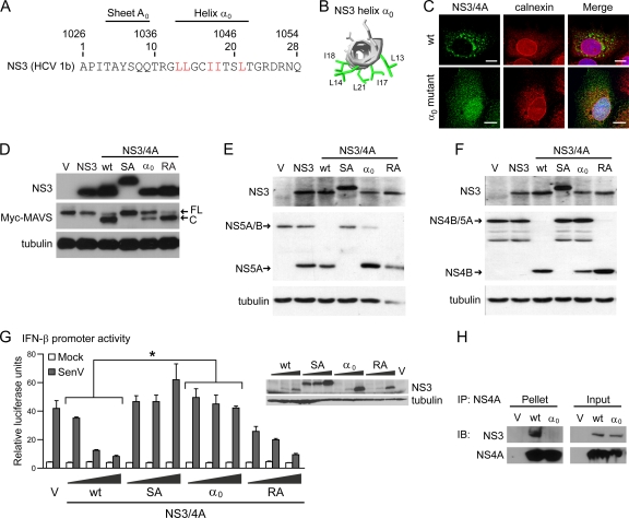
Hepatitis C virus (HCV) infection is sensed in the host cell by the cytosolic pathogen recognition receptor RIG-I. RIG-I signaling is propagated through its signaling adaptor protein MAVS to drive activation of innate immunity. However, HCV blocks RIG-I signaling through viral NS3/4A protease cleavage of MAVS on the mitochondrion-associated endoplasmic reticulum (ER) membrane (MAM). The multifunctional HCV NS3/4A serine protease is associated with intracellular membranes, including the MAM, through membrane-targeting domains within NS4A and also at the amphipathic helix α(0) of NS3. The serine protease domain of NS3 is required for both cleavage of MAVS, a tail-anchored membrane protein, and processing the HCV polyprotein. Here, we show that hydrophobic amino acids in the NS3 helix α(0) are required for selective cleavage of membrane-anchored portions of the HCV polyprotein and for cleavage of MAVS for control of RIG-I pathway signaling of innate immunity. Further, we found that the hydrophobic composition of NS3 helix α(0) is essential to establish HCV replication and infection. Alanine substitution of individual hydrophobic amino acids in the NS3 helix α(0) impaired HCV RNA replication in cells with a functional RIG-I pathway, but viral RNA replication was rescued in cells lacking RIG-I signaling. Therefore, the hydrophobic amphipathic helix α(0) of NS3 is required for NS3/4A control of RIG-I signaling and HCV replication by directing the membrane targeting of both viral and cellular substrates.


Figures

Fig 1
Fig 1
Helix α0 of HCV NS3 is required for MAVS cleavage and RIG-I pathway control by HCV NS3/4A. (A) Diagram of the sequence of the amino terminus of NS3 (HCV Con1 clone; GenBank accession no. AJ238799). Amino acids are numbered with respect to NS3 and the HCV polyprotein (top row). Functional domains are labeled. The hydrophobic amino acids in the NS3 helix α0 are shown in red. (B) Ribbon representation of NS3 aa 12 to 23 using the coordinates provided by Yao et al. (37) (PDB entry 1CU1) with the side chains of the surface-exposed hydrophobic amino acids depicted and labeled. (C) Confocal micrographs of Huh7 cells expressing wt NS3/4A or NS3/4A with mutation of the hydrophobic amino acids in the helix α0 to alanine (α0 mutant) that were immunostained with anti-NS3 (green) and anti-calnexin (red). Nuclei were stained with Draq5 (blue). Bar: 10 μm. (D to F) Immunoblot analysis of extracts from cells expressing the indicated NS3/4A constructs and Myc-MAVS (D), Flag-NS5A/B (E), or Flag-NS4B/5A (F). Arrows mark full-length (FL) and cleaved (C) MAVS in panel D and full-length and cleaved HCV polyprotein in panels E and F. The constructs used express either NS3, wt NS3/4A, NS3/4A S139A (SA), NS3/4A α0 mutant (α0), or NS3/4A R24/26A (RA). (G) IFN-β promoter reporter luciferase expression of Huh7 cells transfected with increasing amounts of NS3/4A expression plasmids and then mock or SenV infected. Values are mean ± standard deviation (SD) (n = 3). *, P ≤ 0.003 by unpaired Student's t test. (Inset) Immunoblot for NS3 protein expression from this assay. (H) Immunoblot analysis of anti-NS3 or anti-NS4A immunoprecipitated extracts, as well as input, from cells expressing wt NS3/4A, α0 mutant NS3/4A, or empty vector (V).
Fig 2
Fig 2
Helix α0 positions the HCV NS3 protease active site at intracellular membranes for substrate cleavage. (A) Confocal micrographs of Huh7 cells expressing wt SC-protease or SC-protease α0 mutant that were immunostained with anti-Flag (SC-protease; green) and anti-calnexin (red). Nuclei were stained with Draq5 (blue). Bar: 10 μm. (B to D) Immunoblot analysis of extracts from cells expressing SC-protease constructs and Myc-MAVS (B), Flag-NS5A/B (C), or Flag-NS4B/5A (D). Arrows mark full-length (FL) and cleaved (C) MAVS in panel B and full-length and cleaved HCV polyprotein in panels C and D. In panel B, the asterisk indicates an alternate form of MAVS likely modified by phosphorylation (32). In panel D, the asterisk marks an alternate SC-protease cleavage product of NS4B/5A.
Fig 3
Fig 3
SC-protease cleaves MAVS at an alternate site to block RIG-I pathway signaling. (A) IFN-β promoter reporter luciferase expression of 293 cells transfected with increasing amounts of SC-protease expression plasmids and then mock or SenV infected. Values are mean ± SD (n = 3) of one of four replicate experiments. (Inset) Immunoblot for Flag-SC-protease protein expression from this assay. (B) Immunoblot analysis of anti-Myc immunoprecipitations and input from 293 cells expressing Flag-MAVS, Myc-MAVS, and increasing amounts of indicated SC-protease constructs (wt, S139A, or α0) or vector. (C) Entire panel of the Flag-MAVS immunoblot from panel B is shown. Arrows mark full-length (FL), cleaved (C; at C508), and alternate cleaved (C-2) forms of MAVS. The asterisk indicates an alternate form of MAVS not resulting from cleavage. (D) The sequence of the HCV polyprotein cleavage sites, as well as known and possible cleavage sites of MAVS, are shown, with the P1 and P1′ residues shown in boldface. (E) Immunoblot analysis of MAVS cleavage at alternate sites by the SC-protease. Blots were probed with anti-Myc (for MAVS) and anti-Flag (for SC-protease). Arrows mark full-length (FL) and cleaved (C, at C508; C-2, at C283) MAVS. (F) IFN-β promoter reporter luciferase expression of 293 cells following MAVS wt overexpression alone or with increasing doses of MAVS aa 284 to 540. Values are mean ± SD (n = 3).
Fig 4
Fig 4
Helix α0 of NS3 is required for RNA replication of HCV subgenomic replicon. (A) RNA was in vitro transcribed from the parental HCV-HP replicon construct (wt), HP replicon harboring the NS3 helix α0 mutations (α0), or individual NS3 amino acid mutations and electroporated into Huh7 and Huh7.5 cells, followed by plating on 100-mm-diameter dishes in serial dilutions, (2 × 10⁵, ×104, and ×103 cells) and crystal violet staining after 3 weeks of G418 selection. The relative transduction efficiency (mean ± SD, from 2 experiments) denotes the percent colonies in Huh7 cells relative to wt as compared those in Huh7.5 cells. (B) A single colony was expanded from Huh7.5 cells replicating the HP replicon with the NS3 helix α0 mutations (referred to as α0REV). Extracts from Huh7.5 cells harboring the wt, the α0REV, or no HP replicon (−) were assayed by immunoblotting for expression of MAVS and HCV proteins (using patient antisera). Arrows mark full-length (FL) and cleaved (C) MAVS; small arrows mark HCV proteins. (C) The hydrophobic amino acids in wt NS3 helix α0 are underlined (top) and the introduced NS3 helix α0 mutations are in bold (middle). The sequence of the α0 revertant (bottom) shows reversion to the parental NS3 amino acids except at L14A. (D and E) Immunoblot analysis of extracts from 293 cells expressing the indicated NS3 constructs and HCV NS5A/B (D) or NS4B/5A (E) using anti-NS3 and anti-Flag (for Flag-tagged HCV polyprotein substrates). Arrows mark full-length and cleaved HCV polyprotein.

References

    1. Baril M, Racine ME, Penin F, Lamarre D. 2009. MAVS dimer is a crucial signaling component of innate immunity and the target of hepatitis C virus NS3/4A protease. J. Virol. 83:1299–1311 - PMC - PubMed
    1. Bartenschlager R, Ahlborn-Laake L, Mous J, Jacobsen H. 1994. Kinetic and structural analyses of hepatitis C virus polyprotein processing. J. Virol. 68:5045–5055 - PMC - PubMed
    1. Beran RK, Pyle AM. 2008. Hepatitis C viral NS3-4A protease activity is enhanced by the NS3 helicase. J. Biol. Chem. 283:29929–29937 - PMC - PubMed
    1. Brass V, et al. 2008. Structural determinants for membrane association and dynamic organization of the hepatitis C virus NS3-4A complex. Proc. Natl. Acad. Sci. U. S. A. 105:14545–14550 - PMC - PubMed
    1. Brenndorfer ED, et al. 2009. Nonstructural 3/4A protease of hepatitis C virus activates epithelial growth factor-induced signal transduction by cleavage of the T-cell protein tyrosine phosphatase. Hepatology 49:1810–1820 - PubMed

Publication types

MeSH terms

Substances