. 2012 Mar;86(6):3152-66.
doi: 10.1128/JVI.06886-11. Epub 2012 Jan 11.

Restricted replication of xenotropic murine leukemia virus-related virus in pigtailed macaques

Affiliations

Restricted replication of xenotropic murine leukemia virus-related virus in pigtailed macaques

Gregory Q Del Prete et al. J Virol. 2012 Mar.

Abstract

Although xenotropic murine leukemia virus-related virus (XMRV) has been previously linked to prostate cancer and myalgic encephalomyelitis/chronic fatigue syndrome, recent data indicate that results interpreted as evidence of human XMRV infection reflect laboratory contamination rather than authentic in vivo infection. Nevertheless, XMRV is a retrovirus of undefined pathogenic potential that is able to replicate in human cells. Here we describe a comprehensive analysis of two male pigtailed macaques (Macaca nemestrina) experimentally infected with XMRV. Following intravenous inoculation with >10(10) RNA copy equivalents of XMRV, viral replication was limited and transient, peaking at ≤2,200 viral RNA (vRNA) copies/ml plasma and becoming undetectable by 4 weeks postinfection, though viral DNA (vDNA) in peripheral blood mononuclear cells remained detectable through 119 days of follow-up. Similarly, vRNA was not detectable in lymph nodes by in situ hybridization despite detectable vDNA. Sequencing of cell-associated vDNA revealed extensive G-to-A hypermutation, suggestive of APOBEC-mediated viral restriction. Consistent with limited viral replication, we found transient upregulation of type I interferon responses that returned to baseline by 2 weeks postinfection, no detectable cellular immune responses, and limited or no spread to prostate tissue. Antibody responses, including neutralizing antibodies, however, were detectable by 2 weeks postinfection and maintained throughout the study. Both animals were healthy for the duration of follow-up. These findings indicate that XMRV replication and spread were limited in pigtailed macaques, predominantly by APOBEC-mediated hypermutation. Given that human APOBEC proteins restrict XMRV infection in vitro, human XMRV infection, if it occurred, would be expected to be characterized by similarly limited viral replication and spread.


Figures
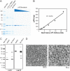
Fig 1
Fig 1
XMRV stock characterization. XMRV-containing supernatants collected from the human prostate carcinoma line 22Rv1 were purified through a sucrose pad and concentrated 1,000× for protein analysis. (A) Total protein in lysed XMRV virions was assessed by SDS-PAGE and Coomassie blue staining. A p30 standard curve for densitometry analysis was established by running serial dilutions of HPLC-purified Mo-MLV p30 protein. Volumes of 0.25 and 0.125 μl of concentrated XMRV were analyzed to confirm that the samples were within the linear range of the standard curve. (B) A densitometry standard curve established using the p30CA standards in panel A was used for linear regression analysis of the p30CA content of the XMRV stock. (C) Western blot analysis of concentrated XMRV using anti-Mo-MLV monoclonal antibodies directed to p30CA (left panel) and gp70SU (right panel). (D) Transmission electron micrographs of 1,000×-concentrated XMRV material at low (left) and high (right) magnifications. MW Stds, molecular weight standards.
Fig 2
Fig 2
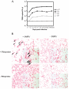
XMRV replication in pigtailed macaque PBMC. (A) Pigtailed macaque PBMC were isolated from whole blood, stimulated with αCD3/IL-2, and infected 3 days poststimulation with 1010, 109, 108, or 106 RNA copy equivalents of 22Rv1-produced XMRV. The vRNA copy number in 8.4 μl of culture supernatant was measured by qRT-PCR at the indicated time points with a sensitivity cutoff of 100 copies. PBMC from three macaques were infected with nearly identical results; data shown are from a representative animal. (B) ISH on in vitro-infected pigtailed macaque PBMC. Pigtailed macaque PBMC were spotted onto slides at 15 dpi and probed using a cocktail of riboprobes directed at antisense gag, pol, pro, and env RNAs. Each panel shows a representative field at 400× and 1,000× (inset) magnifications. Arrows indicate XMRV RNA+ cells. Top left, XMRV-infected cells stained with riboprobes; top right, uninfected cells stained with riboprobes; bottom left, infected cells stained with secondary antibody only with no riboprobes; bottom right, uninfected cells stained with secondary antibody only with no riboprobes.
Fig 3
Fig 3
XMRV replication in pigtailed macaques. vRNA in plasma and vDNA in PBMC, LN, spleen, and prostate tissues were quantified by X-SCA qPCR and qRT-PCR for two XMRV-infected pigtailed macaques, 8242 (A) and 14232 (B). Plasma RNA, PBMC DNA, and peripheral LN DNA were sampled longitudinally at the indicated time points; Bron-LN, Mes-LN, spleen, and prostate tissues were sampled at necropsy at 119 dpi. Plasma RNA values shown are numbers of copies per milliliter; DNA values shown are numbers of copies per million cells.
Fig 4
Fig 4
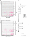
Sequencing analysis of plasma RNA and PBMC DNA from infected macaques. (A and B) Highlighter plots of XMRV gag sequences for single genomes in plasma (RNA) and PBMC (DNA) sampled at the indicated days from animals 8242 (A) and 14232 (B). Individual nucleotide polymorphisms relative to the consensus sequence are indicated by colored ticks (thymine in red, guanine in orange, adenine in green, cytosine in blue, gaps in gray, and guanine-to-adenine changes consistent with APOBEC-mediated hypermutation in pink). (C) Neighbor-joining tree of plasma RNA sequences from animal 8242 rooted on the predominant clone in the 22Rv1-derived XMRV inoculum.
Fig 5
Fig 5
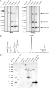
Serum antibody responses in XMRV-infected macaques. (A) Immunoblot assay of 100 ng p30-containing whole, lysed XMRV loaded in each lane and probed with serum collected at the indicated time points diluted 1:2,000. (B) Chromatogram of HPLC-fractionated purified XMRV. Protein sequencing results and the identified protein constituents for each major peak are indicated. Amino acid residues in bold are sites that distinguish XMRV from Mo-MLV. (C) Immunoblot assay of HPLC-fractionated XMRV using macaque serum diluted 1:2,000. A 10-μg sample of p30-containing virus was fractionated, and 50% of each HPLC peak was loaded per lane. Data shown are for week 7 serum from animal 14232. MW Stds, molecular weight standards.
Fig 6
Fig 6
Immunoblot assay titers of serum antibody responses at necropsy. Serial dilutions of serum samples collected at necropsy from animals 8242 (A) and 14232 (B) were used to probe blots with 100 ng p30-containing whole, lysed XMRV loaded per lane. MW Stds, molecular weight standards.
Fig 7
Fig 7
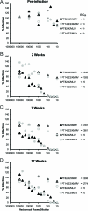
Plasma neutralization activity. Neutralization of 293T-produced XMRV clone VP62 or a 293T-produced, VSV-G-pseudotyped Mo-MLV by heat-inactivated plasma samples collected preinfection (A) and at weeks 2 (B), 7 (C), and 17 (D) postinfection was determined in a multicycle assay on DERSE.LiG-puro indicator cells read at 4 dpi. Percent infection was calculated using the percentage of GFP-positive cells normalized to infection in the absence of macaque plasma. ED50 values represent the reciprocal plasma dilutions at which the percentage of GFP reporter cells was reduced by 50%.
Fig 8
Fig 8
Innate immune response in peripheral LNs. Representative high-magnification (×200) images of T cell zone from peripheral LNs sampled at the indicated time points and immunohistochemically stained for the type I IFN-responsive gene for MxA. MxA staining of an LN from a chronically SIV-infected macaque is shown as a positive control.
Fig 9
Fig 9
CD8+ T cell activation. Quantitative analysis of the percentage of CD8+ Tcm cells (CD95+ CD28+) expressing Ki67 at the indicated time points in blood and LNs of animals 8242 (A) and 14232 (B). The week zero time point represents the day of XMRV inoculation.

References

    1. Akgul B, et al. 6 September 2011. No evidence for a role of xenotropic murine leukaemia virus-related virus and BK virus in prostate cancer of German patients. Med. Microbiol. Immunol. [Epub ahead of print.] doi:10.1007/s00430-011-0215-0 - DOI - PubMed
    1. Aloia AL, et al. 2010. XMRV: a new virus in prostate cancer? Cancer Res. 70:10028–10033 - PMC - PubMed
    1. Arnold RS, et al. 2010. XMRV infection in patients with prostate cancer: novel serologic assay and correlation with PCR and FISH. Urology 75:755–761 - PubMed
    1. Balada E, Castro-Marrero J, Felip L, Vilardell-Tarres M, Ordi-Ros J. 2011. Xenotropic murine leukemia virus-related virus (XMRV) in patients with systemic lupus erythematosus. J. Clin. Immunol. 31:584–587 - PubMed
    1. Barnes E, et al. 2010. Failure to detect xenotropic murine leukemia virus-related virus in blood of individuals at high risk of blood-borne viral infections. J. Infect. Dis. 202:1482–1485 - PubMed

Publication types

MeSH terms

Substances