. 2012 Mar;86(6):2911-8.
doi: 10.1128/JVI.06075-11. Epub 2012 Jan 11.

Rapid and massive virus-specific plasmablast responses during acute dengue virus infection in humans

Affiliations

Rapid and massive virus-specific plasmablast responses during acute dengue virus infection in humans

Jens Wrammert et al. J Virol. 2012 Mar.

Abstract

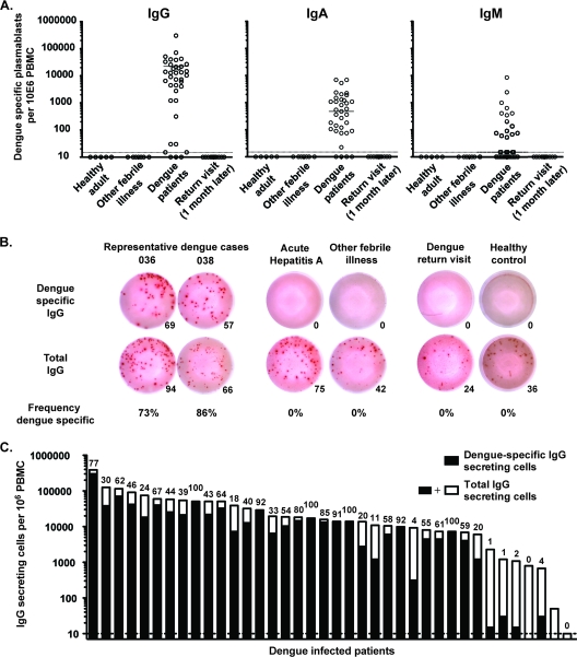
Humoral immune responses are thought to play a major role in dengue virus-induced immunopathology; however, little is known about the plasmablasts producing these antibodies during an ongoing infection. Herein we present an analysis of plasmablast responses in patients with acute dengue virus infection. We found very potent plasmablast responses that often increased more than 1,000-fold over the baseline levels in healthy volunteers. In many patients, these responses made up as much 30% of the peripheral lymphocyte population. These responses were largely dengue virus specific and almost entirely made up of IgG-secreting cells, and plasmablasts reached very high numbers at a time after fever onset that generally coincided with the window where the most serious dengue virus-induced pathology is observed. The presence of these large, rapid, and virus-specific plasmablast responses raises the question as to whether these cells might have a role in dengue immunopathology during the ongoing infection. These findings clearly illustrate the need for a detailed understanding of the repertoire and specificity of the antibodies that these plasmablasts produce.


Figures

Fig 1
Fig 1
Potent plasmablast responses induced during acute dengue virus infection. Plasmablast responses in peripheral blood of healthy controls or patients diagnosed with dengue fever (retrospectively confirmed to be dengue) were measured by flow cytometry. A subset of the patients came back for testing of a follow-up blood sample 1 month after discharge. (A) Representative flow cytometric analysis of the plasmablast frequency in four patients during ongoing dengue virus infection and 1 month after resolution. Plasmablasts are defined herein as CD19+ CD3 CD20−/low CD38hi CD27hi cells. All plots shown are gated on CD3 CD20−/low lymphocytes, including blasting cells. The numbers in the plots represent the percentage of that gate, while numbers in parentheses are the percentage of all lymphocytes. (B) Summary of the frequency of plasmablasts, as a fraction of total CD19+ B cells, in samples obtained from healthy adults, patients with ongoing dengue virus infection, and dengue patients returning after at least 1 month after clearance of infection. Statistical analysis was performed using an unpaired, two-tailed t test. (C) Magnitude of the plasmablast response as absolute numbers in patients infected with different dengue viral serotypes. Statistical analysis was done using an unpaired, two-tailed t test. ND, not detected; ns, not significant. (D) Comparison of the magnitude of the dengue virus infection-induced plasmablast responses to that observed after vaccination with either the inactivated influenza virus vaccine (which induces a recall response that peaks at 7 days after vaccination [35]) or the live attenuated yellow fever virus (YFV) vaccine (which induces a primary response that peaks at day 11/14 after vaccination). Statistical analysis was done using an unpaired, two-tailed t test.
Fig 2
Fig 2
Rapid expansion of plasmablast responses during acute dengue virus infection. (A) Absolute plasmablast numbers per ml blood (calculated using a bead-based [TruCount; BD] system) for all the donors analyzed as a function of number of days after fever onset. Spearman's correlation coefficient test was used to analyze the correlation observed between different parameters for all the data points. Similar results were obtained when each subgroup was analyzed individually. (B) Plasmablast responses in peripheral blood in three patients diagnosed with and confirmed to have dengue fever infection were measured by flow cytometry at two consecutive time points with 2 days between sample collections. Plots show cells gated for CD3 CD20−/low cells with the CD27hi CD38hi plasmablasts marked by a box. The frequency of these plasmablasts is shown next to the box, and the frequency of total lymphocytes is shown in parentheses.
Fig 3
Fig 3
The majority of the plasmablasts induced by dengue virus infection are virus specific. Plasmablast responses in peripheral blood were measured by ELISPOT analysis during infection and 1 month after discharge. (A) Graphs showing the number of IgG-, IgA-, or IgM-secreting dengue virus-specific plasmablasts per million PBMCs. ELISPOT assay plates were coated with purified 16681 DENV-2 antigen. (B) Representative IgG-specific ELISPOT analysis of two dengue virus-infected patients, two patients with other acute febrile illnesses, a dengue patient returning for a 1-month follow-up, and a healthy control. Upper row, dengue virus-specific IgG spots; lower row, total IgG-secreting cells. From left to right, the numbers of cells plated was 823, 823, 2,489, 7,407, 66,000, and 66,000 PBMCs. The number given below each well is the spot count for that well. Relative frequency (percent) of dengue virus-specific IgG-secreting plasmablasts over total IgG secreting plasmablasts is also shown. (C) Summary of the number of total IgG-secreting cells and dengue virus-specific IgG-secreting cells for all the donors analyzed. The entire bar (black and white together) represents the total number of IgG-secreting cells per million PBMCs, while the numbers of dengue virus-specific IgG-secreting cells are marked in black only. Shown above each bar is the percentage of dengue virus-specific cells over total IgG-secreting cells.
Fig 4
Fig 4
Dengue virus-specific serum antibody correlates with the observed plasmablast responses. ELISA analysis of serum samples obtained from the acutely infected dengue patients as well as healthy control samples. This group of samples was from 17 cases of secondary infection, 2 primary cases (based on IgG/IgM ratios, as described in the text), and 3 healthy Thai and 2 healthy U.S. volunteers. Dengue virus-specific IgG was measured by direct ELISA using the same dengue virus antigen used for the ELISPOT analysis of the cellular responses described in the legend to Fig. 3. (A) ELISA measurement of total dengue virus-specific IgG. Midpoint dilution values were determined as the intersection of the dotted line at 50% of the OD value and the ELISA binding curves. (B and C) Correlation of the serological responses (as determined by the midpoint dilution value determined in panel A) and the plasmablast response by absolute plasmablasts in blood (flow cytometry) (B) and dengue virus-specific plasmablasts by ELISPOT assay (C).

References

    1. Akondy RS, et al. 2009. The yellow fever virus vaccine induces a broad and polyfunctional human memory CD8+ T cell response. J. Immunol. 183:7919–7930 - PMC - PubMed
    1. Beltramello M, et al. 2010. The human immune response to dengue virus is dominated by highly cross-reactive antibodies endowed with neutralizing and enhancing activity. Cell Host Microbe 8:271–283 - PMC - PubMed
    1. Bernasconi NL, Traggiai E, Lanzavecchia A. 2002. Maintenance of serological memory by polyclonal activation of human memory B cells. Science 298:2199–2202 - PubMed
    1. Bethell DB, et al. 1998. Pathophysiologic and prognostic role of cytokines in dengue hemorrhagic fever. J. Infect. Dis. 177:778–782 - PubMed
    1. Bhamarapravati N, Tuchinda P, Boonyapaknavik V. 1967. Pathology of Thailand haemorrhagic fever: a study of 100 autopsy cases. Ann. Trop. Med. Parasitol. 61:500–510 - PubMed

Publication types

Substances