. 2012 Apr 5;119(14):3342-51.
doi: 10.1182/blood-2011-11-389668. Epub 2012 Jan 11.

Aβ delays fibrin clot lysis by altering fibrin structure and attenuating plasminogen binding to fibrin

Affiliations

Aβ delays fibrin clot lysis by altering fibrin structure and attenuating plasminogen binding to fibrin

Daria Zamolodchikov et al. Blood. .

Abstract

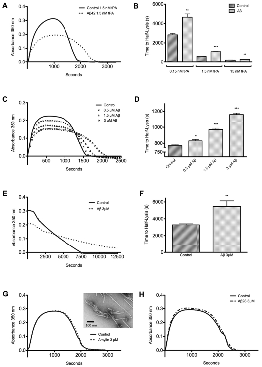
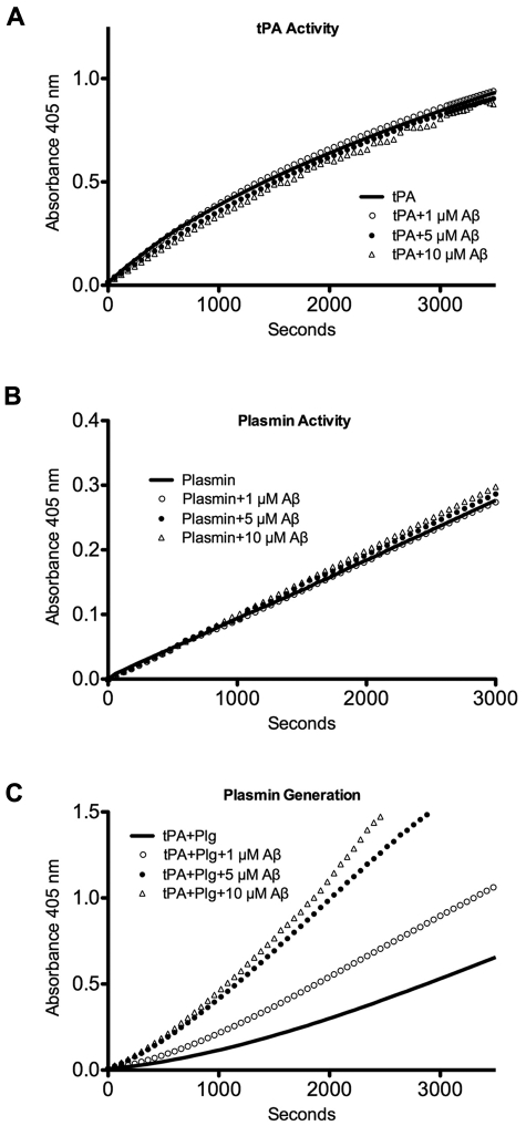
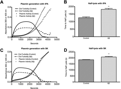
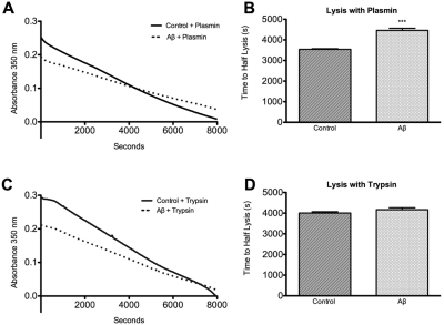
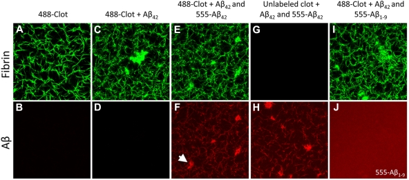
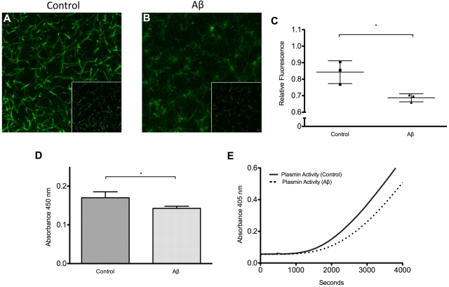
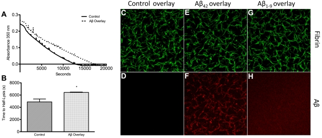
Alzheimer disease is characterized by the presence of increased levels of the β-amyloid peptide (Aβ) in the brain parenchyma and cerebral blood vessels. This accumulated Aβ can bind to fibrin(ogen) and render fibrin clots more resistant to degradation. Here, we demonstrate that Aβ(42) specifically binds to fibrin and induces a tighter fibrin network characterized by thinner fibers and increased resistance to lysis. However, Aβ(42)-induced structural changes cannot be the sole mechanism of delayed lysis because Aβ overlaid on normal preformed clots also binds to fibrin and delays lysis without altering clot structure. In this regard, we show that Aβ interferes with the binding of plasminogen to fibrin, which could impair plasmin generation and fibrin degradation. Indeed, plasmin generation by tissue plasminogen activator (tPA), but not streptokinase, is slowed in fibrin clots containing Aβ(42), and clot lysis by plasmin, but not trypsin, is delayed. Notably, plasmin and tPA activities, as well as tPA-dependent generation of plasmin in solution, are not decreased in the presence of Aβ(42). Our results indicate the existence of 2 mechanisms of Aβ(42) involvement in delayed fibrinolysis: (1) through the induction of a tighter fibrin network composed of thinner fibers, and (2) through inhibition of plasmin(ogen)-fibrin binding.


Figures

Figure 1
Figure 1
Clot lysis is delayed through a range of tPA concentrations in the presence of Aβ42 in a dose-dependent manner. Clot formation and lysis were monitored by turbidity assay. (A) Clot formation and lysis were initiated as described in “Clot turbidity analysis” by combining thrombin, fibrinogen, CaCl2, plasminogen, and 0.15nM, 1.5nM, or 15nM tPA with or without 3μM Aβ42 (0.15nM and 15nM curves not shown because of scale differences). (B) Half-lysis of Aβ clots was significantly longer than for control clots for all concentrations of tPA. (C) Clots were formed as in panel A with 0.5μM, 1.5μM, 3μM Aβ42, or vehicle and 1.5nM tPA. (D) Half-lysis of Aβ clots was significantly delayed in a dose-dependent manner. (E) Preformed clots prepared as described in “Clot turbidity analysis” were overlaid with 50nM tPA. (F) Half-lysis of Aβ clots was significantly longer than control. (G) Clotting and lysis of clots formed as in panel A but with 3μM amylin confirmed by TEM to be fibrillar (inset) did not differ from control. (H) Clots formed as in panel A but with 3μM Aβ1-28 did not differ from control clots (turbidity plots represent mean of 3 experiments; bar graphs represent mean ± SD of 3 experiments; statistical significance noted as *P < .05, **P < .005, and ***P < .0005).
Figure 2
Figure 2
Aβ does not directly inhibit tPA activity, plasmin activity, or plasmin generation from plasminogen.42 at 1μM, 5μM, and 10μM or vehicle was combined with (A) tPA and S-2288 to monitor tPA activity; (B) plasmin and Pefa-5329 to monitor plasmin activity; or (C) tPA, plasminogen (Plg), and Pefa-5329 to monitor plasmin generation from plasminogen. Representative results from ≥ 3 separate experiments.
Figure 3
Figure 3
Plasmin generation by tPA, but not SK, is decreased in Aβ-influenced clots during clotting and lysis. (A) Fibrinogen, plasminogen, tPA, thrombin, CaCl2, and Pefa-5329 or vehicle were mixed with or without 3μM Aβ42 as described in “Enzyme activity.” Absorbance was measured at 350 nm to follow clot formation and lysis and at 405 nm to monitor plasmin activity. Curves of A405 without Pefa-5329 were subtracted from Pefa-5329 A405 curves to control for A405 arising from clot turbidity and not plasmin activity. (B) Half-lysis of tPA/plasminogen-lysed clots was delayed in the presence of Aβ compared with control (P = .011). (C) Same as panel A, except SK was substituted for tPA. (D) Half-lysis of SK/plasminogen-lysed clots was delayed in the presence of Aβ compared with control (P = .005).
Figure 4
Figure 4
Aβ-influenced clots are resistant to lysis by plasmin but not trypsin. (A) Preformed clots prepared as described in “Clot turbidity analysis” with or without 5μM Aβ42 were overlaid with 250nM plasmin. (B) Half-lysis was significantly slower in clots containing Aβ (P = .0001). (C) Preformed clots as in panel A were overlaid with 1μM trypsin. (D) There was no significant difference between half-lysis times of control and Aβ clots (P = .12).
Figure 5
Figure 5
Confocal microscopy of fibrin clots show Aβ binding to fibrin fibrils. Fibrin clots were formed with or without Aβ42 as described in “Aβ binding to fibrin(ogen)” to determine the location of Aβ binding. (Top row) Fibrin visualized with Alexa Fluor 488–labeled fibrinogen (green); (bottom row) Aβ visualized with HiLyte Fluor 555–labeled Aβ (red); (A-B) control clot. (C-D) Clot with only unlabeled Aβ42 shows that fibrin fibers and irregular clusters do not produce signal in the red channel. (E-F) Clot formed with unlabeled Aβ42 and HiLyte Fluor 555–labeled Aβ42 shows colocalization between Aβ and fibrin fibers as well as Aβ and irregular clusters (arrowhead). (G-H) Clot formed without Alexa Fluor 488–labeled fibrinogen but with both unlabeled and labeled Aβ42 shows Aβ signal in the fibrin fiber pattern, confirming that Aβ signal is not Alexa Fluor 488 signal detected in the red channel. (I-J) Clot formed with unlabeled Aβ42 and HiLyte Fluor-555–labeled Aβ1-9 does not have Aβ signal along fibrin fibers or in aggregates, indicating that the Aβ42 signal represents specific Aβ-fibrin(ogen) binding and not fluorophore entrapment. Images are representative of ≥ 3 experiments.
Figure 6
Figure 6
Plasminogen binding to fibrin and plasmin generation is inhibited by Aβ. Fibrin clots were formed with FITC-plasminogen as described in “Plasminogen binding to fibrin,” and 5 μm z-stacks composed of 11 sections were acquired and projected 2-dimensionally for control (A) and Aβ42-containing (B) clots. Images of 15 random sections from 3 separate clots were also acquired and used for quantification (insets show representative single sections). (A) Control clot has formed with FITC-plasminogen shows plasminogen fluorescence in the pattern of the fibrin network. (B) Clot formed with Aβ has less FITC-plasminogen fluorescence. (C) Fluorescence intensity relative to maximum intensity recorded was significantly lower (P = .02) for Aβ-containing clots. (D) Plasminogen binding to fibrin monolayers exposed to 2μM Aβ42 or vehicle was measured by ELISA and normalized to samples not containing plasminogen. Plasminogen binding was decreased in the presence of Aβ (P = .04). (E) Plasmin generation was measured by overlaying tPA, plasminogen, and chromogenic substrate Pefa-5329 on fibrin monolayers exposed to 2μM Aβ42 or vehicle and recording absorbance at 405 nm. Plasmin generation on fibrin monolayers exposed to Aβ was attenuated.
Figure 7
Figure 7
Preformed clots overlaid with Aβ are resistant to lysis and contain fibrin-bound Aβ. (A) Preformed clots (as described in “Clot turbidity analysis”) containing no Aβ were overlaid with 5μM Aβ42 (dashed line) or control buffer (solid line) for 1 hour, the overlays removed, and the clot surfaces washed. All clots were then overlaid with 10nM tPA to initiate lysis. (B) Half-lysis of clots that had been overlaid with Aβ was significantly delayed compared with control clots (P = .012). (C-H) Confocal microscopy of clots using Alexa Fluor 488–labeled fibrinogen and HiLyte Fluor-555–labeled Aβ prepared as described in “Aβ binding to fibrinogen.” (C-D) Normal clot overlaid with buffer. (E-F) Normal clot overlaid with 555-Aβ42 (3μM) for 1 hour contained fibrin-bound Aβ42 (G-H). Normal clot overlaid with 555-Aβ1-9 (3μM) for 1 hour did not show specific colocalization between fibrin and Aβ1-9. Images are representative of ≥ 3 experiments.

Comment in

References

    1. de la Torre JC. Alzheimer disease as a vascular disorder: nosological evidence. Stroke. 2002;33(4):1152–1162. - PubMed
    1. de la Torre JC. Is Alzheimer's disease a neurodegenerative or a vascular disorder? Data, dogma, and dialectics. Lancet Neurol. 2004;3(3):184–190. - PubMed
    1. Smith EE, Greenberg SM. Beta-amyloid, blood vessels, and brain function. Stroke. 2009;40(7):2601–2606. - PMC - PubMed
    1. Iadecola C. Neurovascular regulation in the normal brain and in Alzheimer's disease. Nat Rev Neurosci. 2004;5(5):347–360. - PubMed
    1. Ahn HJ, Zamolodchikov D, Cortes-Canteli M, Norris EH, Glickman JF, Strickland S. Alzheimer's disease peptide beta-amyloid interacts with fibrinogen and induces its oligomerization. Proc Natl Acad Sci U S A. 2010;107(50):21812–21817. - PMC - PubMed

Publication types

MeSH terms