. 2012 Jan 11;4(116):116ra5.
doi: 10.1126/scitranslmed.3002796.

Magnetic resonance of 2-hydroxyglutarate in IDH1-mutated low-grade gliomas

Affiliations

Magnetic resonance of 2-hydroxyglutarate in IDH1-mutated low-grade gliomas

Adam Elkhaled et al. Sci Transl Med. .

Abstract

Recent studies have indicated that a significant survival advantage is conferred to patients with gliomas whose lesions harbor mutations in the genes isocitrate dehydrogenase 1 and 2 (IDH1/2). IDH1/2 mutations result in aberrant enzymatic production of the potential oncometabolite D-2-hydroxyglutarate (2HG). Here, we report on the ex vivo detection of 2HG in IDH1-mutated tissue samples from patients with recurrent low-grade gliomas using the nuclear magnetic resonance technique of proton high-resolution magic angle spinning spectroscopy. Relative 2HG levels from pathologically confirmed mutant IDH1 tissues correlated with levels of other ex vivo metabolites and histopathology parameters associated with increases in mitotic activity, relative tumor content, and cellularity. Ex vivo spectroscopic measurements of choline-containing species and in vivo magnetic resonance measurements of diffusion parameters were also correlated with 2HG levels. These data provide extensive characterization of mutant IDH1 lesions while confirming the potential diagnostic value of 2HG as a surrogate marker of patient survival. Such information may augment the ability of clinicians to monitor therapeutic response and provide criteria for stratifying patients to specific treatment regimens.


Figures
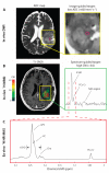
Fig. 1
Fig. 1
Designating tissue targets from presurgical in vivo imaging. (A) ADC map derived from diffusion-weighted imaging (DWI) shows a hypointense region of low diffusion relative to normal-appearing white matter that was designated as a target for tissue sampling in a WHO grade III astrocytoma. Tissue targets were defined as 5-mm-diameter spheres on surgical navigation software. (B) In vivo MRSI (extent of coverage defined by yellow box) overlaid on T1-weighted spoiled gradient echo (SPGR) image along with a choline-to-NAA index (CNI) color map. A spectrum from a region of elevated CNI targeted for tissue sampling is displayed in the green box. Cr, creatine; NAA, N-acetylaspartate; PCr, phosphocreatine; tCHO, total choline. (C) Sample ex vivo 1H HR-MAS spectrum from tissue acquired at the site of elevated CNI for comparison with in vivo spectroscopy data in (B). Cho, free choline; GPC, glycerophosphocholine; PC, phosphocholine.
Fig. 2
Fig. 2
IDH1R132H immunostaining and direct sequencing of IDH1 gDNA. (A) IDH1R132H antibody staining (brown) on a 2HG+ WHO grade III astrocytoma tissue sample, which was representative of 44 tissue samples. The corresponding 1D and 2D spectra for this sample are shown in Fig. 3, A and C, respectively. Scale bar, 100 μm. (B) Complementary image of a 2HG− sample on which immunostaining for IDH1R132H was also performed and no mutant enzymes were found (representative of seven samples). (C) Electropherogram of polymerase chain reaction (PCR)–amplified gDNA performed on an IDH1R132H-mutant tissue sample. Codon 132 displays signal from the mutant base pair composed of an adenine (mutant) and guanine [wild-type (WT)] nucleobase, each present on complimentary strands of DNA. The heterozygous substitution of adenine at R132 results in the production of histidine at the substrate binding site of IDH1 and neomorphic enzymatic activity.
Fig. 3
Fig. 3
Ex vivo spectra from mutant and WT IDH1 tissue samples. (A and B) 1D CPMG spectrum from an IDH1-mutant WHO grade III astrocytoma (A) and WT IDH1 grade IV astrocytoma (B). For both (A) and (B), the blue trace represents acquired data and the red trace represents the spectral fit used for relative quantification of 2HG levels by HR-QUEST. These spectra are not normalized with respect to each other. (C) 2D TOCSY spectrum of IDH1-mutant tissue depicted in (A). Shared magnetization between nuclei of discrete molecular spin systems is rendered through resonant cross peaks about the F2/F1 diagonal. The correspondence among individual cross peaks of 2HG is highlighted along with the uniquely identifying spectral feature created with respect to the α-proton, labeled as 2HGα. Ace, acetate; Ala, alanine; Gln, glutamine; Glu, glutamate; Gly, glycine; Lac, lactate; myo-I, myo-inositol; scyllo-I, scylloinositol; Tau, taurine.
Fig. 4
Fig. 4
2HG levels according to WHO glioma grade. (A) Plot of relative 2HG levels quantified by HR-QUEST in relation to malignancy grades. Mean relative levels per patient (n = 29 total: 7, grade II; 18, grade III; 4, grade IV) were evaluated by an exact Wilcoxon rank-sum test. Brackets indicate the statistical comparison of grade II 2HG levels with those of gliomas that had transformed to a higher grade; significance was defined as P < 0.05. (B) These levels are also shown normalized with respect to cellularity (number of cells per 200× field) for those patients whose tissue samples could be evaluated for this parameter (n = 22 total: 6, grade II; 13, grade III; 3, grade IV). NS, not significant.

References

    1. Grier JT, Batchelor T. Low-grade gliomas in adults. Oncologist. 2006;11:681–693. - PubMed
    1. Riemenschneider MJ, Reifenberger G. Molecular neuropathology of low-grade gliomas and its clinical impact. Adv. Tech. Stand. Neurosurg. 2010;35:35–64. - PubMed
    1. Yan H, Parsons DW, Jin G, McLendon R, Rasheed BA, Yuan W, Kos I, Batinic-Haberle I, Jones S, Riggins GJ, Friedman H, Friedman A, Reardon D, Herndon J, Kinzler KW, Velculescu VE, Vogelstein B, Bigner DD. IDH1 and IDH2 mutations in gliomas. N. Engl. J. Med. 2009;360:765–773. - PMC - PubMed
    1. Parsons DW, Jones S, Zhang X, Lin JC, Leary RJ, Angenendt P, Mankoo P, Carter H, Siu IM, Gallia GL, Olivi A, McLendon R, Rasheed BA, Keir S, Nikolskaya T, Nikolsky Y, Busam DA, Tekleab H, Diaz LA, Jr., Hartigan J, Smith DR, Strausberg RL, Marie SK, Shinjo SM, Yan H, Riggins GJ, Bigner DD, Karchin R, Papadopoulos N, Parmigiani G, Vogelstein B, Velculescu VE, Kinzler KW. An integrated genomic analysis of human glioblastoma multiforme. Science. 2008;321:1807–1812. - PMC - PubMed
    1. Houillier C, Wang X, Kaloshi G, Mokhtari K, Guillevin R, Laffaire J, Paris S, Boisselier B, Idbaih A, Laigle-Donadey F, Hoang-Xuan K, Sanson M, Delattre JY. IDH1 or IDH2 mutations predict longer survival and response to temozolomide in low-grade gliomas. Neurology. 2010;75:1560–1566. - PubMed

Publication types

MeSH terms