. 2012 Mar;23(5):930-44.
doi: 10.1091/mbc.E11-11-0927. Epub 2012 Jan 11.

Dissection of the NUP107 nuclear pore subcomplex reveals a novel interaction with spindle assembly checkpoint protein MAD1 in Caenorhabditis elegans

Affiliations

Dissection of the NUP107 nuclear pore subcomplex reveals a novel interaction with spindle assembly checkpoint protein MAD1 in Caenorhabditis elegans

Eduardo Ródenas et al. Mol Biol Cell. 2012 Mar.

Abstract

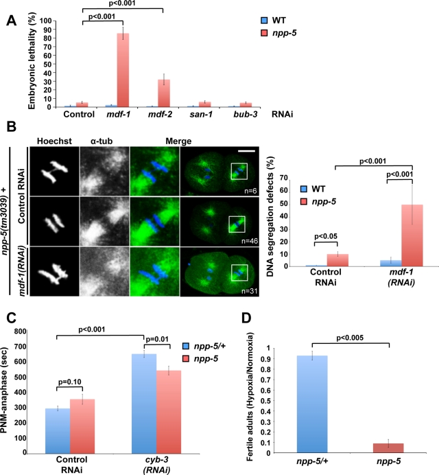
Nuclear pore complexes consist of several subcomplexes. The NUP107 complex is important for nucleocytoplasmic transport, nuclear envelope assembly, and kinetochore function. However, the underlying molecular mechanisms and the roles of individual complex members remain elusive. We report the first description of a genetic disruption of NUP107 in a metazoan. Caenorhabditis elegans NUP107/npp-5 mutants display temperature-dependent lethality. Surprisingly, NPP-5 is dispensable for incorporation of most nucleoporins into nuclear pores and for nuclear protein import. In contrast, NPP-5 is essential for proper kinetochore localization of NUP133/NPP-15, another NUP107 complex member, whereas recruitment of NUP96/NPP-10C and ELYS/MEL-28 is NPP-5 independent. We found that kinetochore protein NUF2/HIM-10 and Aurora B/AIR-2 kinase are less abundant on mitotic chromatin upon NPP-5 depletion. npp-5 mutants are hypersensitive to anoxia, suggesting that the spindle assembly checkpoint (SAC) is compromised. Indeed, NPP-5 interacts genetically and physically with SAC protein MAD1/MDF-1, whose nuclear envelope accumulation requires NPP-5. Thus our results strengthen the emerging connection between nuclear pore proteins and chromosome segregation.


Figures

FIGURE 1:
FIGURE 1:
npp-5(tm3039) and npp-5(ok1966) are null mutations of NUP107/npp-5. (A) Schematic representation of the C. elegans npp-5 gene and deletion alleles npp-5(ok1966) and npp-5(tm3039). Exons and introns are indicated by boxes and lines, respectively. (B) Western blot analysis of ∼1500 wild-type (WT), npp-5(ok1966), and npp-5(tm3039) embryos probed with anti-NPP-5 and anti–α-tubulin antibodies. NPP-5 appeared with a molecular weight of ∼92 kDa in wild-type embryos only. Asterisk indicates nonspecific cross-reactivity. (C) Wild type and npp-5(tm3039) embryos were fixed and stained with anti–NPP-5 antiserum (red) and monoclonal antibody mAb414 (green). Chromatin was detected using Hoechst 33258 (blue). Boxed regions in the merged panels are shown at higher magnification to the left. Scale bar, 10 μm. (D) Percentage of wild-type, npp-5(ok1966) and npp-5(tm3039) offspring dying during embryogenesis (Emb) or larval development (Lvl) or reaching adulthood (Adult) at 20°C. Error bars, SE of the mean.
FIGURE 2:
FIGURE 2:
Nuclear protein import occurs in the absence of NPP-5. (A) Size of P1 nuclei was determined by time-lapse microscopy, revealing a significantly slower nuclear growth rate in npp-5(tm3039) embryos (n = 13) compared with npp-5(tm3039)/+ embryos (n = 12). Time is relative to P0 anaphase onset. (B) Gonads of wild-type (n = 14) and npp-5(tm3039) (n = 16) animals were injected with a mixture of 70- (green) and 155-kDa (magenta) dextrans. Exclusion of the dextrans from embryonic nuclei was observed by live confocal microscopy. (C) Nuclear import of PIE-1-GFP into P2 nuclei was observed by time-lapse microscopy (left) and quantified (right). Import in npp-5(tm3039) embryos (n = 10) was comparable to that in control embryos (n = 10). Time is relative to P1 anaphase onset. Error bars, SE of the mean. Scale bars, 10 μm.
FIGURE 3:
FIGURE 3:
Expression and localization of most Nups are NPP-5 independent. (A) Western blot analysis of embryonic extracts showed similar expression levels of NPP-7, NPP-10N, NPP-10C, and NPP-19 in npp-5(tm3039) embryos compared with the wild type. In contrast, NPP-15 appeared as a duplet in the wild type but not in the mutant. (B–D) Wild type, npp-5(tm3039) (B, D), and npp-5(ok1966) (C) embryos were fixed and stained with serum against NPP-10C (B), MEL-28 (C), or NPP-15 (D) (red) and mAb414 (green). Chromatin was detected using Hoechst 33258 (blue). (E, F) Expression of GFP-NPP-23 and GFP-NPP-2/mCherry-HIS-58 was analyzed by live microscopy. Whereas depletion of NPP-5 inhibited recruitment of GFP-NPP-23 (n ≥ 24 for each treatment; E), GFP-NPP-2 was unaffected (F; n ≥ 8 for each treatment). Boxed regions in the merged panels are shown at higher magnification to the left. Scale bars, 10 μm.
FIGURE 4:
FIGURE 4:
NPP-5 localization is sensitive to perturbations of other NUP107 complex members. Still images from time-lapse confocal microscopy of embryos expressing GFP-NPP-5 (green) and mCherry-HIS-58 (magenta). RNAi against npp-2, npp-10, or npp-6 inhibited proper localization of GFP-NPP-5 during interphase (top rows) and metaphase (bottom rows; n ≥ 8 for each treatment). Boxed regions on the right are shown at higher magnification to the left. Scale bar, 10 μm.
FIGURE 5:
FIGURE 5:
NPP-5 is required for efficient localization of HIM-10 and AIR-2. Still images from time-lapse confocal microscopy of control (npp-5(tm3039)/+) and npp-5(tm3039) embryos expressing GFP-HIM-10 (A), GFP-MIS-12 (B), or GFP-AIR-2 (C). Boxed regions (middle) are shown at higher magnification to the left. Scale bars, 10 μm. Graphs on the right represent mean fluorescence intensities measured in 5 × 2.3 μm2 rectangles perpendicular to the metaphase plates. Position is relative to the center of metaphase chromosomes. Probability values (p) from two-tailed t tests are shown. Error bars, SE of the mean. n ≥ 15 in all experiments.
FIGURE 6:
FIGURE 6:
NPC localization of spindle assembly checkpoint protein MDF-1 depends on NPP-5. (A–C) Still images from time-lapse confocal microscopy of embryos expressing GFP-MDF-1 (green) and mCherry-HIS-58 (magenta). (A) Depletion of NPP-5 induces recruitment of GFP-MDF-1 to metaphase chromosomes (arrow; n = 10). (B) GFP-MDF-1 accumulation on chromosomes attached to monopolar spindles in zyg-1(RNAi) embryos is unaffected by depletion of NPP-5 (n = 3). (C) Localization of GFP-MDF-1 to NPCs in interphase is abolished in the absence of NPP-5 (n = 14). (D) Wild-type and npp-5(tm3039) embryos were fixed and stained with anti–MDF-1 antiserum (red) and mAb414 (green). Chromatin was detected using Hoechst 33258 (blue). Boxed regions in the merged panels are shown at higher magnification to the left. Scale bars, 10 μm.
FIGURE 7:
FIGURE 7:
NPP-5 physically interacts with MDF-1 in yeast-two-hybrid assays. Full-length npp-5 and mdf-1 cDNAs were cloned into prey and bait vectors, respectively, and used to transform yeast cells. Growth on selective (–Trp-Leu-His-Ade) medium was only supported when the two genes were present together. No interaction was observed between NPP-19 and MDF-1.
FIGURE 8:
FIGURE 8:
NPP-5 mutants are hypersensitive to MDF-1 depletion and anoxic stress. (A) Embryonic lethality was measured following RNAi against different members of the SAC in wild-type and npp-5(tm3039) animals (n > 600 embryos in all experiments). (B) Wild-type and npp-5(tm3039) animals treated with control or MDF-1 RNAi were fixed and stained with anti–α-tubulin (green). Chromatin was detected using Hoechst 33258 (blue). Boxed regions in the merged panels are shown at higher magnification to the left. Percentages of embryos presenting DNA segregation defects are indicated in the graph, demonstrating a synthetic phenotype in embryos simultaneously depleted for NPP-5 and MDF-1. (C) Time from pronuclear meeting (PNM) to anaphase was measured following incubation on control or cyb-3 RNAi plates for 30 h (n ≥ 5 for all treatments). (D) Embryos from npp-5(tm3039)/+ and npp-5(tm3039) animals were exposed to hypoxia for 21 h. After recovery under normoxic conditions development into fertile adults was determined, revealing a severe effect in npp-5(tm3039) mutants (n > 600 embryos in all experiments). Probability values (p) are shown from chi-square tests (A, B), two-tailed t test (C), and Wilcoxon rank sum test (D). Error bars, SE of the mean.

References

    1. Alber F, et al. The molecular architecture of the nuclear pore complex. Nature. 2007;450:695–701. - PubMed
    1. Askjaer P, Galy V, Hannak E, Mattaj IW. Ran GTPase cycle and importins alpha and beta are essential for spindle formation and nuclear envelope assembly in living Caenorhabditis elegans embryos. Mol Biol Cell. 2002;13:4355–4370. - PMC - PubMed
    1. Bai SW, Rouquette J, Umeda M, Faigle W, Loew D, Sazer S, Doye V. The fission yeast Nup107-120 complex functionally interacts with the small GTPase Ran/Spi1 and is required for mRNA export, nuclear pore distribution, and proper cell division. Mol Cell Biol. 2004;24:6379–6392. - PMC - PubMed
    1. Belgareh N, et al. An evolutionarily conserved NPC subcomplex, which redistributes in part to kinetochores in mammalian cells. J Cell Biol. 2001;154:1147–1160. - PMC - PubMed
    1. Boehmer T, Enninga J, Dales S, Blobel G, Zhong H. Depletion of a single nucleoporin, Nup107, prevents the assembly of a subset of nucleoporins into the nuclear pore complex. Proc Natl Acad Sci USA. 2003;100:981–985. - PMC - PubMed

Publication types

MeSH terms