. 2012 Mar;23(5):781-91.
doi: 10.1091/mbc.E11-06-0537. Epub 2012 Jan 11.

Matrix rigidity regulates a switch between TGF-β1-induced apoptosis and epithelial-mesenchymal transition

Affiliations

Matrix rigidity regulates a switch between TGF-β1-induced apoptosis and epithelial-mesenchymal transition

Jennifer L Leight et al. Mol Biol Cell. 2012 Mar.

Abstract

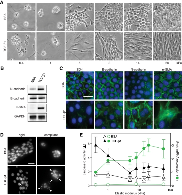
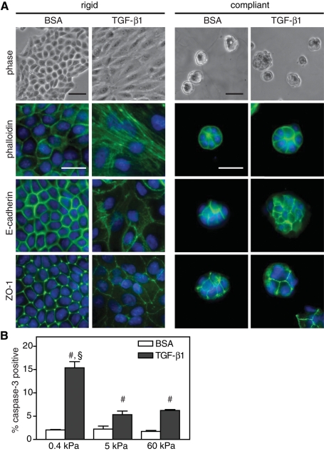
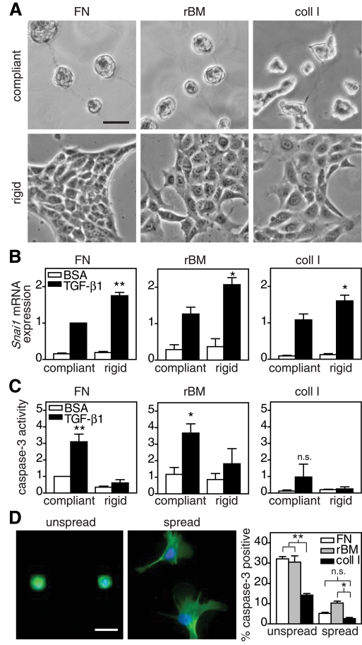
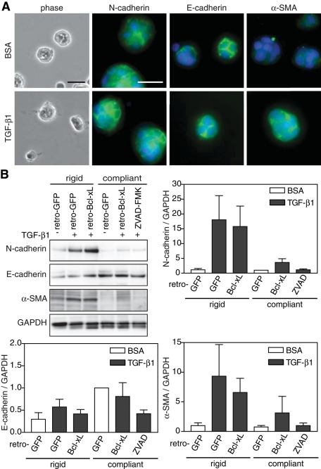
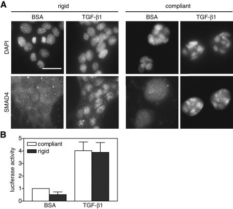
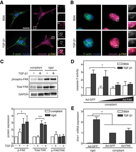
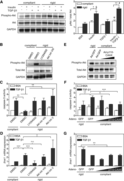
The transforming growth factor-β (TGF-β) signaling pathway is often misregulated during cancer progression. In early stages of tumorigenesis, TGF-β acts as a tumor suppressor by inhibiting proliferation and inducing apoptosis. However, as the disease progresses, TGF-β switches to promote tumorigenic cell functions, such as epithelial-mesenchymal transition (EMT) and increased cell motility. Dramatic changes in the cellular microenvironment are also correlated with tumor progression, including an increase in tissue stiffness. However, it is unknown whether these changes in tissue stiffness can regulate the effects of TGF-β. To this end, we examined normal murine mammary gland cells and Madin-Darby canine kidney epithelial cells cultured on polyacrylamide gels with varying rigidity and treated with TGF-β1. Varying matrix rigidity switched the functional response to TGF-β1. Decreasing rigidity increased TGF-β1-induced apoptosis, whereas increasing rigidity resulted in EMT. Matrix rigidity did not change Smad signaling, but instead regulated the PI3K/Akt signaling pathway. Direct genetic and pharmacologic manipulations further demonstrated a role for PI3K/Akt signaling in the apoptotic and EMT responses. These findings demonstrate that matrix rigidity regulates a previously undescribed switch in TGF-β-induced cell functions and provide insight into how changes in tissue mechanics during disease might contribute to the cellular response to TGF-β.


Figures

FIGURE 1:
FIGURE 1:
Matrix rigidity regulates TGF-β1–induced EMT and apoptosis in NMuMG cells. (A) Phase contrast images of cells cultured on PA gels with elastic modulus ranging from 0.4 to 60 kPa and treated with TGF-β1 or BSA control. (B) Western blot of N-cadherin (135 kDa), E-cadherin (120 kDa), α-SMA (42 kDa), and GAPDH control (38 kDa) in cells cultured on rigid (8 kPa) PA gels. (C) Immunofluorescence images of cells cultured on rigid PA gels. (D) Hoechst-stained nuclei of cells cultured on rigid (8 kPa) and compliant (0.4 kPa) gels. Fragmented nuclei indicated by white triangles. (E) Caspase-3 activity (▲, ∆) and Snai1 mRNA expression (•, ○) in cells cultured on PA gels. n = 5 ± SEM. Bars, 50 μm.
FIGURE 2:
FIGURE 2:
TGF-β1–induced EMT and apoptosis in MDCK cells cultured on polyacrylamide gels. (A) Immunostaining on rigid (5 kPa) and compliant (0.4 kPa) PA gels in cells treated with TGF-β1 or BSA control. (B) Graph of percentage of cells positive for cleaved caspase-3 immunofluorescence. n = 3 ± SEM. #p < 0.01 as compared with BSA conditions; §p < 0.01 as compared with 5- and 60-kPa TGF-β1 conditions. Bars, 50 μm.
FIGURE 3:
FIGURE 3:
TGF-β1–induced EMT and apoptosis in NMuMG cells cultured on polyacrylamide gels conjugated with ECM. (A) Phase contrast images of cells cultured on FN, rBM, or coll I conjugated PA gels. (B) Snai1 mRNA expression in cells on compliant and rigid gels treated with TGF-β1. (C) Caspase-3 activity in cells on compliant and rigid gels treated with TGF-β1. (D) Phalloidin (green)- and Hoechst (blue)-stained cells on 289 μm2 islands (unspread) or large areas (spread) of microcontact-printed FN. Graph of percentage of cells treated with TGF-β1 positive for cleaved caspase-3 immunofluorescence. n = 3 ± SEM. *p < 0.05; **p < 0.01. Bars, 50 μm.
FIGURE 4:
FIGURE 4:
Decreased matrix rigidity inhibits EMT independent of apoptosis. (A) Phase contrast and immunostaining for N-cadherin, E-cadherin, α-SMA, and nuclei of NMuMG cells infected with retro-Bcl-xL on compliant gels. (B) Western blot and quantification of N-cadherin, E-cadherin, α-SMA, and GAPDH in NMuMG cells infected with retro-GFP or retro-Bcl-xL or treated with 400 μM ZVAD-FMK, plated on rigid and compliant gels, and treated with TGF-β1. n = 4 ± SEM. Bars, 50 μm.
FIGURE 5:
FIGURE 5:
Smad signaling in NMuMG cells on compliant and rigid gels. (A) Immunostaining for Smad4 and nuclei in NMuMG cells cultured on rigid and compliant PA gels treated with TGF-β1. (B) Luciferase activity in cells transfected with 3TP-luciferase reporter plasmid on compliant and rigid gels. n = 4 ± SEM. Bars, 50 μm.
FIGURE 6:
FIGURE 6:
Effect of matrix rigidity and TGF-β1 on the actin cytoskeleton and focal adhesion formation in NMuMG cells. (A,B) Immunofluorescence images of F-actin (green), nuclei (blue), vinculin (magenta), and phospho-FAK (yellow) on rigid (8 kPa) (A) and compliant (0.4 kPa) (B) gels. Inset shows magnification of vinculin (V), phospho-FAK (F), and merged (M) images. (C) Western blot and quantification of phospho-FAK (125 kDa), total FAK (125 kDa), and GAPDH (38 kDa). Caspase-3 activity (D) and Snai1 mRNA expression (E) in NMuMG cells infected with Ad-GFP or Ad-FAK cultured on rigid and compliant gels treated with TGF-β1. n = 4 ± SEM. *p < 0.05; **p < 0.01; ***p < 0.001. Bars, 25 μm.
FIGURE 7:
FIGURE 7:
Regulation of Akt activity by matrix rigidity in NMuMG cells. (A) Western blot and quantification of phospho-Akt (60 kDa), total Akt (60 kDa), and GAPDH (38 kDa) in cells plated on compliant and rigid polyacrylamide gels. (B–D) Caspase-3 activity and Snai1 mRNA expression in cells treated with DMSO control, 10 μM LY294002, 1 μM Akt inhibitor VIII, or 10 μM Akt inhibitor V. (E–G) Caspase-3 activity and Snai1 mRNA expression in cells infected with Ad-GFP or Ad-p110-CAAX. n = 3 ± SEM. *p < 0.05; **p < 0.01; ***p < 0.001; §p < 0.01 rigid + insulin compared with BSA and TGF-β1 conditions; #p < 0.01 rigid + insulin/TGF-β1 compared with all conditions; φp < 0.01 compliant + insulin/TGF-β1 compared with compliant BSA condition.

References

    1. Alcaraz J, Xu R, Mori H, Nelson CM, Mroue R, Spencer VA, Brownfield D, Radisky DC, Bustamante C, Bissell MJ. Laminin and biomimetic extracellular elasticity enhance functional differentiation in mammary epithelia. EMBO J. 2008;27:2829–2838. - PMC - PubMed
    1. Arora PD, Narani N, McCulloch CA. The compliance of collagen gels regulates transforming growth factor-beta induction of alpha-smooth muscle actin in fibroblasts. Am J Pathol. 1999;154:871–882. - PMC - PubMed
    1. Bacman D, Merkel S, Croner R, Papadopoulos T, Brueckl W, Dimmler A. TGF-beta receptor 2 downregulation in tumour-associated stroma worsens prognosis and high-grade tumours show more tumour-associated macrophages and lower TGF-beta1 expression in colon carcinoma: a retrospective study. BMC Cancer. 2007;7:156. - PMC - PubMed
    1. Bailey KM, Liu J. Caveolin-1 up-regulation during epithelial to mesenchymal transition is mediated by focal adhesion kinase. J Biol Chem. 2008;283:13714–13724. - PMC - PubMed
    1. Bakin AV, Tomlinson AK, Bhowmick NA, Moses HL, Arteaga CL. Phosphatidylinositol 3-kinase function is required for transforming growth factor beta-mediated epithelial to mesenchymal transition and cell migration. J Biol Chem. 2000;275:36803–36810. - PubMed

Publication types

MeSH terms

Substances