. 2012 Mar;23(5):800-10.
doi: 10.1091/mbc.E11-11-0950. Epub 2012 Jan 11.

Derlin-1 and UBXD8 are engaged in dislocation and degradation of lipidated ApoB-100 at lipid droplets

Affiliations

Derlin-1 and UBXD8 are engaged in dislocation and degradation of lipidated ApoB-100 at lipid droplets

Michitaka Suzuki et al. Mol Biol Cell. 2012 Mar.

Abstract

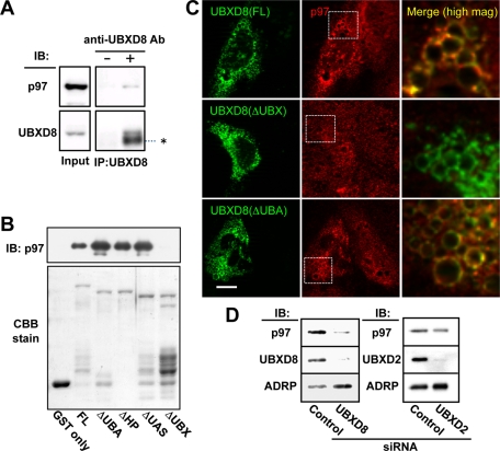
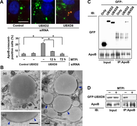
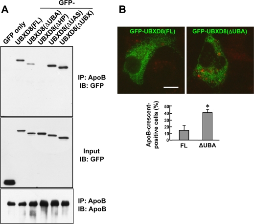
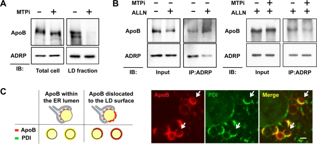
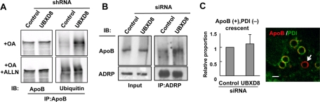
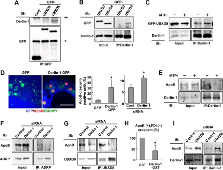
Apolipoprotein B-100 (ApoB) is the principal component of very low density lipoprotein. Poorly lipidated nascent ApoB is extracted from the Sec61 translocon and degraded by proteasomes. ApoB lipidated in the endoplasmic reticulum (ER) lumen is also subjected to proteasomal degradation, but where and how it dislocates to the cytoplasm remain unknown. In the present study, we demonstrate that ApoB after lipidation is dislocated to the cytoplasmic surface of lipid droplets (LDs) and accumulates as ubiquitinated ApoB in Huh7 cells. Depletion of UBXD8, which is almost confined to LDs in this cell type, decreases recruitment of p97 to LDs and causes an increase of both ubiquitinated ApoB on the LD surface and lipidated ApoB in the ER lumen. In contrast, abrogation of Derlin-1 function induces an accumulation of lipidated ApoB in the ER lumen but does not increase ubiquitinated ApoB on the LD surface. UBXD8 and Derlin-1 bind with each other and with lipidated ApoB and show colocalization around LDs. These results indicate that ApoB after lipidation is dislocated from the ER lumen to the LD surface for proteasomal degradation and that Derlin-1 and UBXD8 are engaged in the predislocation and postdislocation steps, respectively.


Figures

FIGURE 1:
FIGURE 1:
UBXD8 and other ERAD-related proteins were present in LDs of Huh7 cells. The cells were maintained in the normal culture medium without any fatty acid supplement in this and subsequent experiments unless otherwise stated. (A) Fractions obtained by sucrose density-gradient ultracentrifugation were analyzed by Western blotting. LDs were recovered in the fraction of the lowest density (fraction 1), which was verified by enrichment of ADRP. In addition to UBXD8, UBXD2, and p97, which were identified by mass spectrometric analysis, Derlin-1 and SEL1L were also found in the LD fraction. In contrast, Sec61α was found only in the bottom fractions (fractions 6–8), in which most membrane and soluble proteins were contained. SEL1L was immunoprecipitated before SDS–PAGE for efficient detection. (B) Immunofluorescence microscopy confirmed that UBXD8, UBXD2, and p97 distributed in LDs. LDs and nuclei were labeled with BODIPY493/503 (green) and 4′,6-diamidino-2-phenylindole (blue), respectively. p97 distributed throughout the cytoplasm and nucleoplasm, but a conspicuous concentration around LDs was observed. Bars, 10 μm. (C) Double immunofluorescence labeling revealed that UBXD8 (red) and an ER luminal protein, PDI (green), were distributed on the opposite hemisphere of LDs. Bar, 5 μm. (D) Domain structure and hydropathy plot of UBXD8. A domain showing high hydrophobicity was named the HP domain. Full-length UBXD8 and mutants lacking one of the domains (ΔUBA, ΔHP, ΔUAS, or ΔUBX) were conjugated to the carboxy terminus of GFP and expressed in Huh7 cells. (E) GFP-UBXD8(ΔUBA), GFP-UBXD8(ΔUAS), GFP-UBXD8(ΔUBX), and GFP-HP showed concentration around LDs like GFP-UBXD8(FL), but GFP alone and GFP-UBXD8(ΔHP) distributed diffusely in the cytoplasm, indicating that the HP domain is necessary and sufficient for UBXD8 to localize in the LD. LDs were stained red by BODIPY558/568-C12. Bars, 10 μm.
FIGURE 2:
FIGURE 2:
UBXD8 recruited p97 to LDs by the UBX domain. (A) Immunoprecipitation of endogenous UBXD8 from Huh7 cell lysates caused cosedimentation of endogenous p97. The input lane was loaded with 2% of the cell lysate. The IgG heavy chain is indicated by an asterisk. (B) In vitro pull-down assay between recombinant p97 and recombinant GST-UBXD8 proteins. An equal amount of GST-free p97 was incubated with full-length and mutant GST-UBXD8 proteins, and the bound fraction was examined by Western blotting. All but GST and GST-UBXD8(ΔUBX) pulled down p97. The GST proteins in the input are shown by Coomassie brilliant blue staining. (C) Endogenous UBXD8 in Huh7 cells was knocked down by siRNA, and siRNA-resistant cDNA of GFP-tagged UBXD8(FL), UBXD8(ΔUBX), or UBXD8(ΔUBA) was transfected. In cells expressing the GFP-tagged proteins (green), LDs that harbored GFP-UBXD8(FL) and UBXD8(ΔUBA) showed an intense accumulation of endogenous p97 (red), but those with UBXD8(ΔUBX) did not. Merged pictures show a high magnification of the rectangular areas. Bar, 10 μm. (D) Huh7 cells were depleted of UBXD8 or UBXD2 by RNAi and the amount of p97 in the LD fraction was compared. p97 in the LD fraction was reduced significantly after UBXD8 knockdown but less so by UBXD2 knockdown. ADRP is shown as a loading control of LDs.
FIGURE 3:
FIGURE 3:
Knockdown of UBXD8 increased ApoB-crescents. (A) Huh7 cells were depleted of either UBXD8 or UBXD2, and the proportion of cells bearing ApoB-crescents was determined after labeling ApoB (red), LDs (green), and nuclei (blue). ApoB in the bulk ER lumen was not labeled when cells were permeabilized with digitonin (Ohsaki et al., 2008). Knockdown of UBXD8, but not that of UBXD2, significantly increased the number of ApoB-crescents in Huh7 cells (Student's t test; *p < 0.01). The increase of ApoB-crescents in cells depleted of UBXD8 was suppressed by treating cells with 100 nM BAY13-9952 (MTPi) for 12 or 72 h before fixation. The result is representative of three independent experiments. Bar, 10 μm. (B) Huh7 cells transfected with control siRNA (a) or UBXD8 siRNA (b, c) were observed by electron microscopy. ApoB-crescents made of an LD and a thin cistern fusing to it (arrowheads) were observed frequently in cells depleted of UBXD8. Bars, 500 nm. (C) Huh7 cells expressing GFP-UBXD8 or GFP-UBXD2 were treated with 10 μM acetyl-leucinyl-leucinyl-norleucinal (ALLN) for 12 h, lysed, and immunoprecipitated with anti-ApoB antibody. GFP-UBXD8, but not GFP-UBXD2, coprecipitated with ApoB. The ALLN treatment was carried out to increase ubiquitinated ApoB, but essentially the same result was obtained without the treatment. (D) Huh7 cells were treated with 10 μM ALLN alone or with 10 μM ALLN and 100 nM BAY13-9952 for 12 h before lysis and immunoprecipitation. GFP-UBXD8 showed coimmunoprecipitation with ApoB, but its amount was reduced drastically when MTP was inhibited.
FIGURE 4:
FIGURE 4:
The UBA domain of UBXD8 is important for degradation of lipidated ApoB. (A) Huh7 cells expressing GFP-tagged UBXD8 proteins were treated with 10 μM ALLN for 12 h, lysed, and immunoprecipitated with anti-ApoB antibody. GFP was used as a negative control. GFP-UBXD8(ΔUBA) and GFP-UBXD8(ΔHP) showed significantly less coprecipitation with ApoB than the others. (B) Huh7 cells were transfected with cDNAs of GFP-UBXD8(FL) or GFP-UBXD8(ΔUBA) and labeled for ApoB (red). The proportion of cells bearing ApoB-crescents was considerably higher in cells expressing GFP-UBXD8(ΔUBA) than in those expressing GFP-UBXD8(FL). The result shown in the bar graph is representative of three independent experiments. Bar, 10 μm.
FIGURE 5:
FIGURE 5:
ApoB after lipidation was detected on the cytoplasmic surface of LDs. (A) Huh7 cells were treated with or without 100 nM BAY13-9952 for 48 h, and ApoB in the total cell lysate and the LD fraction was examined. ApoB in the LD fraction was virtually eliminated by the MTPi treatment, whereas the total ApoB in the cell was reduced to only a minor degree. ADRP is shown as a loading control of the LD fraction. (B) Huh7 cells were treated with 10 μM ALLN alone or with 10 μM ALLN and 100 nM BAY13-9952 for 12 h, and then reacted with 1 mM DSP, a membrane-permeable cross-linker, for 30 min on ice before lysis. ApoB cross-linkable with ADRP increased by proteasomal inhibition (left), but the increase was suppressed when MTP was inhibited simultaneously (right). (C) Huh7 cells were treated with 10 μM ALLN for 12 h and labeled for ApoB (red) and PDI (green). The diagram shows the rationale that dislocated ApoB should give rise to ApoB-positive, PDI-negative labeling on the entire or partial LD surface. Confocal microscopy showed that ApoB is present on the surface devoid of PDI in some ApoB-crescents (arrows). Bar, 1 μm.
FIGURE 6:
FIGURE 6:
Knockdown of UBXD8 increased ApoB on the cytoplasmic surface of LDs. (A) Huh7 cells stably harboring control or UBXD8 shRNA were incubated with or without 10 μM ALLN for 12 h, and the LD fractions were solubilized and immunoprecipitated with anti-ApoB antibody. OA (0.4 mM) was added to the culture medium to increase LDs. ApoB in the LD fraction after UBXD8 knockdown showed increased ubiquitination, and the intensity was comparable to that induced by ALLN. (B) Huh7 cells transfected with control or UBXD8 siRNA were treated with 1 mM DSP for cross-linking. ApoB cross-linkable to ADRP was increased significantly by UBXD8 depletion. (C) The dislocation assay by immunofluorescence microscopy. Huh7 cells were transfected with control or UBXD8 siRNA, treated with 10 μM ALLN for 12 h, and labeled for ApoB (red) and PDI (green). For unambiguous quantification, the proportion of entirely red labels among ApoB-crescents was compared (arrow). The result in the bar graph is the average of three independent experiments. The ApoB-positive, PDI-negative labeling persisted after UBXD8 knockdown, indicating that UBXD8 is dispensable for the ApoB dislocation. Bar, 1 μm.
FIGURE 7:
FIGURE 7:
Inhibition of Derlin-1 function suppressed the cytoplasmic dislocation of ApoB. (A) Huh7 cells expressing GFP, GFP-UBXD2, or GFP-UBXD8 were lysed and immunoprecipitated by anti-GFP antibody. Endogenous Derlin-1 coprecipitated with GFP-UBXD8 significantly, whereas it bound with no GFP or little GFP-UBXD2. The IgG heavy and light chains are indicated by asterisk and double asterisks, respectively. (B) Huh7 cells expressing GFP-UBXD2 or GFP-UBXD8 were lysed and immunoprecipitated by anti–Derlin-1 antibody. GFP-UBXD8 coimmunoprecipitated significantly with endogenous Derlin-1, whereas little GFP-UBXD2 did so. (C) Huh7 cells treated with 10 μM ALLN alone or with 10 μM ALLN and 100 nM BAY13-9952 for 12 h were lysed, and endogenous Derlin-1 was immunoprecipitated. Comparable amounts of GFP-UBXD8 coprecipitated with Derlin-1 irrespective of the MTPi treatment. (D) Huh7 cells expressing GFP or dominant-negative Derlin-1 (Derlin-1–GFP; green) were labeled for ApoB (red) and LDs (blue). Bar, 10 μm. The frequency of ApoB-crescent–positive cells was significantly higher in cells expressing Derlin-1–GFP than in those expressing GFP. ApoB-crescents also increased by Derlin-1 knockdown. The average of results from three independent experiments is shown (Student's t test; *p < 0.05). (E) Huh7 cells were treated as in (C). ApoB coprecipitating with Derlin-1 was reduced significantly by the MTPi treatment for 12 h. (F) Huh7 cells transfected with control or Derlin-1 siRNA were incubated with 10 μM ALLN for 12 h. ApoB cross-linkable to ADRP with 1 mM DSP was reduced by Derlin-1 knockdown. (G) Huh7 cells were treated as in (F). ApoB coimmunoprecipitating with UBXD8 was reduced by Derlin-1 knockdown. (H) The dislocation assay by immunofluorescence microscopy. Huh7 cells were transfected with cDNA of either GST alone or Derlin-1–GST, treated with 10 μM ALLN for 12 h, and labeled for ApoB (red), PDI (green), and GST (blue). Derlin-1–GST was used instead of Derlin-1–GFP to employ the same fluorophore combination for ApoB and PDI as in Figures 5C and 6C. The proportion of ApoB+, PDI spheres was significantly lower in Derlin-1-GST–positive cells than that in the control, indicating that the dominant-negative Derlin-1 abrogated the cytoplasmic dislocation of ApoB. The average of three independent experiments is shown. (I) Huh7 cells were transfected with control or UBXD8 siRNA. UBXD8 knockdown did not influence the amount of ApoB coimmunoprecipitating with Derlin-1.
FIGURE 8:
FIGURE 8:
Derlin-1 and UBXD8 were distributed in proximity around LDs. (A) In situ PLA to identify the location where two endogenous proteins exist in proximity. The approximation signal (red), LDs (green), and nuclei (blue) are shown. The number of the approximation signal was much higher for the combination of Sec61α and Sec61β (30.7/cell) than for the combination of Derlin-1 and UBXD8 (3.4/cell), but the proportion of the approximation signal associated with LDs was significantly higher for Derlin-1–UBXD8 than for Sec61α-Sec61β (Student's t test; *p < 0.05). The relative LD area was equivalent in the two samples (Derlin-1–UBXD8, 2.8%; Sec61α-Sec61β, 2.6%). The average of results from three independent experiments is shown. Negative control using the combination of anti–Derlin-1 antibody and nonimmune goat IgG did not give any approximation signal. Bar, 10 μm. (B) Schematic diagram of UBXD8, Derlin-1, and p97 interactions at the LD intercalated in the ER membrane. Only lipidated ApoB in the ER lumen is depicted here, but the present data indicated that some ApoB after lipidation is dislocated and present on the cytoplasmic surface of the LD.

References

    1. Alexandru G, Graumann J, Smith GT, Kolawa NJ, Fang R, Deshaies RJ. UBXD7 binds multiple ubiquitin ligases and implicates p97 in HIF1alpha turnover. Cell. 2008;134:804–816. - PMC - PubMed
    1. Bagola K, Mehnert M, Jarosch E, Sommer T. Protein dislocation from the ER. Biochim Biophys Acta. 2011;1808:925–936. - PubMed
    1. Bartz R, Zehmer JK, Zhu M, Chen Y, Serrero G, Zhao Y, Liu P. Dynamic activity of lipid droplets: protein phosphorylation and GTP-mediated protein translocation. J Proteome Res. 2007;6:3256–3265. - PubMed
    1. Bernardi KM, Forster ML, Lencer WI, Tsai B. Derlin-1 facilitates the retro-translocation of cholera toxin. Mol Biol Cell. 2008;19:877–884. - PMC - PubMed
    1. Brasaemle DL, Barber T, Kimmel AR, Londos C. Post-translational regulation of perilipin expression. Stabilization by stored intracellular neutral lipids. J Biol Chem. 1997;272:9378–9387. - PubMed

Publication types