. 2012 May;40(9):4040-51.
doi: 10.1093/nar/gkr1271. Epub 2012 Jan 11.

C7orf30 specifically associates with the large subunit of the mitochondrial ribosome and is involved in translation

Affiliations

C7orf30 specifically associates with the large subunit of the mitochondrial ribosome and is involved in translation

Bas F J Wanschers et al. Nucleic Acids Res. 2012 May.

Abstract

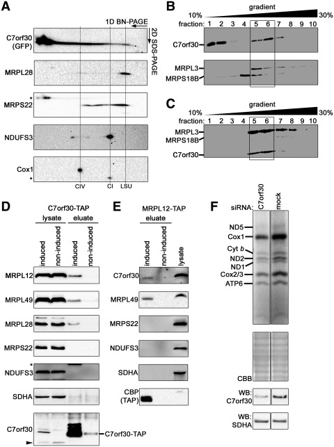
In a comparative genomics study for mitochondrial ribosome-associated proteins, we identified C7orf30, the human homolog of the plant protein iojap. Gene order conservation among bacteria and the observation that iojap orthologs cannot be transferred between bacterial species predict this protein to be associated with the mitochondrial ribosome. Here, we show colocalization of C7orf30 with the large subunit of the mitochondrial ribosome using isokinetic sucrose gradient and 2D Blue Native polyacrylamide gel electrophoresis (BN-PAGE) analysis. We co-purified C7orf30 with proteins of the large subunit, and not with proteins of the small subunit, supporting interaction that is specific to the large mitoribosomal complex. Consistent with this physical association, a mitochondrial translation assay reveals negative effects of C7orf30 siRNA knock-down on mitochondrial gene expression. Based on our data we propose that C7orf30 is involved in ribosomal large subunit function. Sequencing the gene in 35 patients with impaired mitochondrial translation did not reveal disease-causing mutations in C7orf30.


Figures

Figure 1.
Figure 1.
Conservation of the bacterial operon that contains the bacterial ortholog of C7orf30, ybeB. The gene order is conserved in divergent branches of bacteria. Solid lines connect neighboring genes in the genomes of the given clade (right), a dotted line is used if both genes are not direct neighbors, but include other genes in between. rplU, ribosomal protein L21; rpmA, ribosomal protein L27; obgE, GTPase; proB, glutamate 5-kinase; proA, gamma-glutamyl phosphate reductase; nadD, nicotinic acid mononucleotide adenylyltransferase; ybeB, C7orf30 homolog; rlmH, 23S rRNA methyltransferase. Data from the STRING database (39).
Figure 2.
Figure 2.
Domain composition of the human C7orf30 protein. (A) Predicted structure of the conserved DUF143 domain modeled after the Bacillus halodurans ortholog (pdb:2o5a, Benach et al., submitted for publication). Homology model was created with WHAT IF (54) and visualized with PyMOL 1.3 (http://www.pymol.org). Side chains of residues conserved in all orthologs are show in blue. (B) Functional domains of the human C7orf30 protein. N-terminal targeting signal was predicted with Target P (44). Bacterial homologs possess only the DUF143 domain. (C) Multiple alignment and secondary structure of the conserved DUF143 domain. Non-conserved insertions in S. cerevisiae and B. hominis are replaced with dots. Mitochondrial and chloroplast proteins are marked with mt and ch, respectively. The alignment was visualized using Jalview (55) using the Clustalx color scheme. Residues conserved in all orthologs are marked above the alignment. Secondary structure α-helix (red) and β-sheet (yellow) regions are marked beneath the alignment together with their position in the homology model of C7orf30. See Figure 2 for the list of species used in the alignment.
Figure 3.
Figure 3.
Maximum likelihood phylogeny (see ‘Materials and Methods’ section for details) of C7orf30 homologs reveals distinct chloroplast/cyanobacterial and mitochondrial/proteobacterial clusters. Species included: Rickettsia prowazekii, Escherchia coli, Toxoplasma gondii, Blastocystis hominis, Leishmania major, Saccharomyces cerevisiae, Ostreococcus lucimarinus, Dictyostelium discoideum, Schizosaccharomyces pombe, Homo sapiens, Zea mays, Arabidopsis thaliana, Synechococcus sp. PCC 7002 and Cyanothece sp. PCC 7424.
Figure 4.
Figure 4.
C7orf30 is a mitochondrial matrix localized protein absent in cells without mitochondrial DNA. (A) Confocal microscope analysis of HeLa cells immunostained with anti-C7orf30 (left panel) and anti-MRPL12 (middle panel) antibodies. The merged image is shown on the right. Nuclei are counterstained with Hoechst. (B) Cellular fractionation of HEK293 cells. Total cell lysate (TC), cytosolic (Cyt.) and mitochondrial (Mit.)-enriched fractions were analyzed using western blotting with indicated antibodies. As control of mitochondrial and cytoplasmic fractions TOM20 (mitochondrial outer membrane) and anti-CK-B (Creatine kinase B-type) antibodies were used, respectively. The 75-kDa signal visible after C7orf30 antibody incubation presumably corresponds to a cross-reacting contaminant. (C) C7orf30 resides within the boundaries of the mitochondrial inner membrane. Mitochondria with digitonin permeabilized outer membranes were subjected to the amounts of proteinase K indicated, in the absence or presence of membrane dissolving Triton X-100 (TX-100). Susceptibility of proteins to degradation was analyzed with western blotting using indicated antibodies; TOM20: mitochondrial outer membrane, prohibitin 1: intermembrane space facing protein, SDHA: matrix. Quantification of the immunoreactive bands is indicated at the right. (D) C7orf30 is not detectable in cells devoid of mitochondrial DNA. SDS–PAGE analysis of fibroblast Cy73, mitochondrial DNA less (Rho0), HeLa and HEK293 cells. Membranes were probed with the antibodies indicated. Cox1 (mitochondrial encoded) and SDHA (nuclear encoded) were used as controls. (E) Inhibition of mitochondrial translation does not affect C7orf30 protein levels. Mitochondrial translation was inhibited with chloramphenicol (CAP) and allowed to resume again for an increasing amount of time. Protein levels were analyzed with SDS–PAGE followed by western blotting. Immunostainings for Cox2 (mitochondrial encoded) and SDHA were used as controls. Asterisk indicates remaining Cox2 signal.
Figure 5.
Figure 5.
C7orf30 associates with the large subunit of the mitochondrial ribosome and is important for mitochondrial translation. (A) Two-dimensional Blue Native PAGE analysis of C7orf30-GFP. Mitochondrial lysate of HEK293 expressing C7orf30-GFP was subjected to separation in the first and second dimension under native and denaturing conditions, respectively before western blot analysis with indicated antibodies. The vertical lines indicate the position of the large subunit (LSU), complex I (CI) and complex IV (CIV). Asterisk denotes signal from previous incubation. (B) Western blot analysis of an isokinetic sucrose gradient centrifugation (see ‘Materials and Methods’ section) immunostained for C7orf30. To indicate the positions of the mitochondrial ribosomal subunits the membranes were additionally stained for MRPL3 and MRPS18B, representing the large subunit and small subunit respectively (lower panel). Although the two subunits have a similar mass, MRPL3 migrates slightly more slowly on SDS–PAGE and can be seen in fractions 5, 6. (C) C7orf30 co-sediments with the large subunit bound pool of ICT1. HEK293 expressing ICT1-FLAG (large subunit protein) were subjected to immunoprecipitation with FLAG-antibody followed by elution of bound proteins. The entire eluate was separated by isokinetic sucrose gradient centrifugation before Western blot analysis with indicated antibodies. (D and E) Western blot analysis of C7orf30-TAP (D) and MRPL12-TAP (E) affinity purifications. Used antibodies are indicated on the left. As control noninduced cells were used. Efficiency of the C7orf30-TAP pull down was assessed by probing the blots with anti-C7orf30 (lower panel). The arrow indicates the signal for endogenous C7orf30, asterisk the signal from a previous incubation. MRPL12-TAP purification was assessed by probing the blots with anti-CBP antibody recognizing the TAP-tag. SDHA and NDUFS3 were used as control to rule out a-specific protein binding. (F) Metabolic labeling of mitochondrial translation products with 35S in HEK293 cells transfected with siRNAs targeting C7orf30 and the mock control. Mitochondrial translation products are indicated on the left. Equal loading of the gels was confirmed by rehydrating and staining the gel with Coomassie Brilliant Blue G-250 (CBB). Knock-down of proteins was verified by western blot (WB) and probing the membranes with indicated antibodies. SDHA was used as loading control.

Comment on

  • Nucleic Acids Res.

References

    1. Fromont-Racine M, Senger B, Saveanu C, Fasiolo F. Ribosome assembly in eukaryotes. Gene. 2003;313:17–42. - PubMed
    1. Kressler D, Linder P, de La Cruz J. Protein trans-acting factors involved in ribosome biogenesis in Saccharomyces cerevisiae. Mol. Cell. Biol. 1999;19:7897–7912. - PMC - PubMed
    1. Saveanu C, Namane A, Gleizes PE, Lebreton A, Rousselle JC, Noaillac-Depeyre J, Gas N, Jacquier A, Fromont-Racine M. Sequential protein association with nascent 60S ribosomal particles. Mol. Cell. Biol. 2003;23:4449–4460. - PMC - PubMed
    1. Dennerlein S, Rozanska A, Wydro M, Chrzanowska-Lightowlers ZM, Lightowlers RN. Human ERAL1 is a mitochondrial RNA chaperone involved in the assembly of the 28S small mitochondrial ribosomal subunit. Biochem. J. 2010;430:551–558. - PMC - PubMed
    1. Uchiumi T, Ohgaki K, Yagi M, Aoki Y, Sakai A, Matsumoto S, Kang D. ERAL1 is associated with mitochondrial ribosome and elimination of ERAL1 leads to mitochondrial dysfunction and growth retardation. Nucleic Acids Res. 2010;38:5554–5568. - PMC - PubMed

Publication types

MeSH terms