Review
. 2012 Feb;97(2):311-25.
doi: 10.1210/jc.2011-2332. Epub 2012 Jan 11.

Update on bone anabolics in osteoporosis treatment: rationale, current status, and perspectives

Affiliations
Review

Update on bone anabolics in osteoporosis treatment: rationale, current status, and perspectives

Roland Baron et al. J Clin Endocrinol Metab. 2012 Feb.

Abstract

Osteoporosis is defined as low bone mineral density associated with skeletal fractures secondary to minimal or no trauma, most often involving the spine, the hip, and the forearm. The decrease in bone mineral density is the consequence of an unbalanced bone remodeling process, with higher bone resorption than bone formation. Osteoporosis affects predominantly postmenopausal women, but also older men. This chronic disease represents a considerable medical and socioeconomic burden for modern societies. The therapeutic options for the treatment of osteoporosis have so far comprised mostly antiresorptive drugs, in particular bisphosphonates and more recently denosumab, but also calcitonin and, for women, estrogens or selective estrogen receptor modulators. These drugs have limitations, however, in particular the fact that they lead to a low turnover state where bone formation decreases with the decrease in bone-remodeling activity. In this review, we discuss the alternative class of osteoporosis drugs, i.e. bone anabolics, their biology, and the perspectives they offer for our therapeutic armamentarium. We focus on the two main osteoanabolic pathways identified as of today: PTH, the only anabolic drug currently on the market; and activation of canonical Wnt signaling through inhibition of the endogenous inhibitors sclerostin and dickkopf1. Each approach is based on a different molecular mechanism, but most recent evidence suggests that these two pathways may actually converge, at least in part. Whereas recombinant human PTH treatment is being revisited with different formulations and attempts to regulate endogenous PTH secretion via the calcium-sensing receptor, antibodies to sclerostin and dickkopf1 are currently in clinical trials and may prove to be even more efficient at increasing bone mass, possibly independent of bone turnover. Each of these anabolic approaches has its own limitations and safety issues, but the prospects of effective anabolic therapy for osteoporosis are indeed bright.


Figures

Fig. 1.
Fig. 1.
Schematic of the remodeling and modeling activities under physiological conditions, in osteoporosis, and during anabolic treatment. A, Within an active BMU under physiological conditions, bone is constantly removed by osteoclasts (OCs) during the resorption phase of the remodeling cycle. After the reversal phase, new bone matrix is produced by osteoblasts (OBs) during the formation phase at sites where bone resorption has occurred with the amount of bone formed being equal of the amount of bone resorbed, thereby maintaining bone mass. Once the BMU is completed, osteoblasts become entrapped as osteocytes (OCYs) into the newly formed matrix, remain on the bone surface as lining cells (LCs), or undergo apoptosis. Bone then remains in the quiescence phase until a new BMU is initiated. B, In osteoporosis, bone resorption is increased and bone formation is decreased, resulting in a loss of bone. C, Administration of recombinant human PTH (rhPTH) stimulates both osteoclast-mediated bone resorption and osteoblast-mediated bone formation, resulting in a high bone turnover with a net gain in bone mass. In addition to its remodeling-based bone anabolic effect, rhPTH also induces bone formation on surfaces around the resorption sites that were not previously subject to bone resorption (modeling). D, Activation of the canonical Wnt signaling pathway tends to decrease bone resorption but mostly increases both remodeling-based and modeling-based bone formation, thereby causing a striking increase in bone formation, particularly in areas that were not previously resorbed (modeling).
Fig. 2.
Fig. 2.
Effects of the two main anabolic pathways, PTH and Wnt signaling, on osteoblasts, and indirectly on osteoclasts. PTH and Wnt both stimulate the proliferation of mesenchymal stem cells (MSCs) and the commitment of these cells into the osteoblast (OB) lineage, whereas the differentiation into chondrocytes and adipocytes is prevented by canonical Wnt signaling. In the late OB, both pathways increase the mineral apposition and bone formations rate (MAR, BFR). In addition, the Wnt pathway stimulates the production of osteoprotegerin (OPG), a soluble decoy receptor for the RANKL, preventing osteoclast (OC) differentiation and function. In contrast, PTH stimulates the secretion of RANKL, which binds to its receptor RANK on OC precursor cells (OC-pre) and mature OCs, thereby stimulating OC differentiation, function, and ultimately bone resorption. Wnt activity is inhibited by sclerostin and Dkk1, both secreted by late OBs and osteocytes. PTH represses the expression of both sclerostin and Dkk1, whereas Dkk1 expression is increased by Wnt activity, establishing a negative feedback loop. Thus, PTH and Wnt signaling pathways increase bone formation through several mechanisms, but only the Wnt pathway represses bone resorption, whereas PTH stimulates OCs via the induction of RANKL production by OBs.
Fig. 3.
Fig. 3.
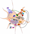
Signaling and cross talk of the PTH and Wnt signaling pathways in osteocytes. In the osteocyte (and late osteoblasts), activation of the canonical Wnt signaling pathway occurs upon simultaneous binding of the secreted glycoprotein Wnt3a (or other Wnts like Wnt 10b for instance) to the seven-helix-receptor frizzled (Fz) family and the coreceptors Lrp 5/6. Binding of Wnt3a to Lrp5/6 changes the conformation of the cytoplasmic receptor domain, causing the recruitment of Axin2 and preventing the phosphorylation of β-catenin by GSK3β and its proteasomal degradation. β-Catenin accumulates in the cytosol and translocates into the nucleus, thereby stimulating the expression of the Lrp5/6 antagonists Dkk1 and sclerostin, and the RANKL inhibitor OPG, via the T-cell factor/lymphoid enhancer factor (Tcf/Lef). PTH binds to its seven-transmembrane-spanning receptor and activates phosphatidyl inositol-specific phospholipase C (PLC), cAMP-dependent protein kinase A (PKA), and the protein kinase C (PKC) downstream signaling cascades, all contributing to the bone anabolic effect of PTH. In addition, PTH signaling cross talks with the Wnt signaling pathway by associating with Lrp6, inhibiting GSK3β, stabilizing β-catenin, and inhibiting the expression of both sclerostin and Dkk1.

References

    1. Parfitt AM, Drezner MK, Glorieux FH, Kanis JA, Malluche H, Meunier PJ, Ott SM, Recker RR. 1987. Bone histomorphometry: standardization of nomenclature, symbols, and units. Report of the ASBMR Histomorphometry Nomenclature Committee. J Bone Miner Res 2:595–610 - PubMed
    1. Bonewald LF. 2011. The amazing osteocyte. J Bone Miner Res 26:229–238 - PMC - PubMed
    1. Harvey N, Dennison E, Cooper C. 2010. Osteoporosis: impact on health and economics. Nat Rev Rheumatol 6:99–105 - PubMed
    1. Hollick RJ, Reid DM. 2011. Role of bisphosphonates in the management of postmenopausal osteoporosis: an update on recent safety anxieties. Menopause Int 17:66–72 - PubMed
    1. Spiro AS, Beil FT, Timo Beil F, Schinke T, Schilling AF, Eulenburg C, Rueger JM, Amling M. 2010. Short-term application of dexamethasone enhances bone morphogenetic protein-7-induced ectopic bone formation in vivo. J Trauma 69:1473–1480 - PubMed

Substances