. 2012 Jan 31;109(5):1548-53.
doi: 10.1073/pnas.1119905109. Epub 2012 Jan 11.

Synthetic in vivo validation of gene network circuitry

Affiliations

Synthetic in vivo validation of gene network circuitry

Sagar S Damle et al. Proc Natl Acad Sci U S A. .

Abstract

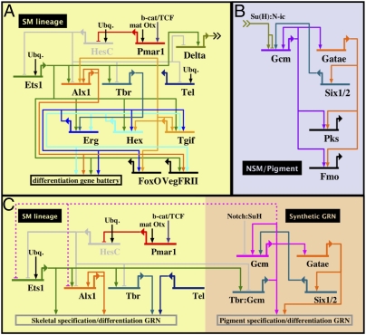
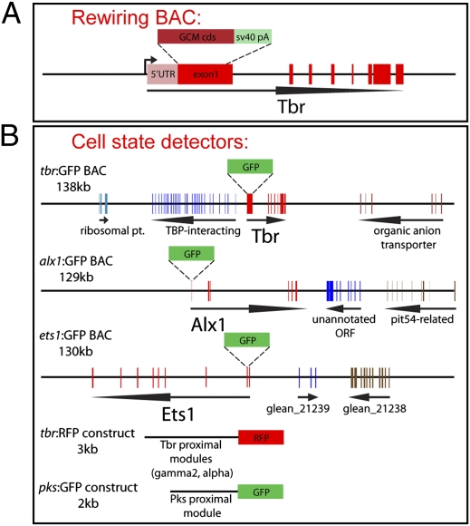
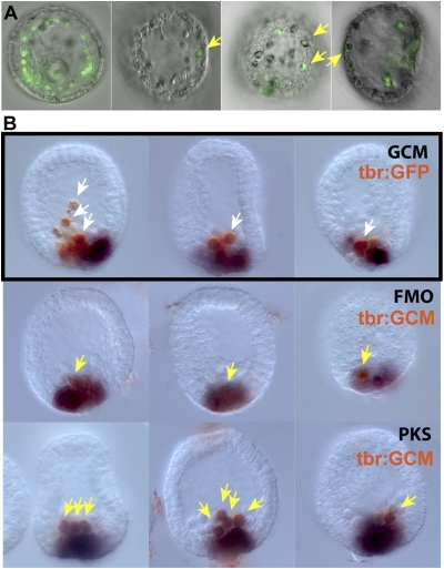
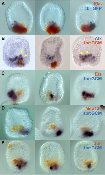
Embryonic development is controlled by networks of interacting regulatory genes. The individual linkages of gene regulatory networks (GRNs) are customarily validated by functional cis-regulatory analysis, but an additional approach to validation is to rewire GRN circuitry to test experimentally predictions derived from network structure. Here we use this synthetic method to challenge specific predictions of the sea urchin embryo endomesoderm GRN. Expression vectors generated by in vitro recombination of exogenous sequences into BACs were used to cause elements of a nonskeletogenic mesoderm GRN to be deployed in skeletogenic cells and to detect their effects. The result of reengineering the regulatory circuitry in this way was to divert the developmental program of these cells from skeletogenesis to pigment cell formation, confirming a direct prediction of the GRN. In addition, the experiment revealed previously undetected cryptic repression functions.


Conflict of interest statement

The authors declare no conflict of interest.

Figures

Fig. 1.
Fig. 1.
Relevant portions of the GRNs for skeletogenic and nonskeletogenic mesoderm specification and differentiation in the sea urchin embryo. (A) Skeletogenic specification, as described in text. Output linkages for the genes tbr and tel are not shown as their targets are off the map. (B) Notch-signaling circuitry leading to activation of gcm and pigment cell differentiation. (C) Diagram of rewired skeletogenic specification network. Gcm expression is brought under control of the double-negative regulatory gate and is expressed in precursors to the skeletogenic mesenchyme. Because the endogenous gcm gene autoactivates, once expressed in skeletogenic cells, the Gcm factor may contribute to maintenance of expression of this factor. Because Gcm is a terminal regulator of the pigment cell differentiation battery, it would be expected to activate this subnetwork in cells originally destined to be skeletogenic. The pink dotted repression bars on Alx1 and Ets1 indicate the possibility, as discussed later in this paper in Cryptic Exclusion of Skeletogenic Regulatory State, of cross-repressive functions downstream of gcm expression that would lower expression of the skeletogenic regulatory state. A full diagram of the endomesodermal gene regulatory network can be found at http://sugp.caltech.edu/endomes/#EndoNetworkDiagrams.
Fig. 2.
Fig. 2.
Diagram of BAC constructs used in the synthetic rewiring experiment. (A) gcm-coding sequence was inserted using homologous recombination into the first exon of the tbr gene in a 140-kb BAC that contains the entire tbr regulatory architecture. (B) A similar knock-in strategy was used to generate BAC-GFP reporters that were used as detectors for measuring cell state. BAC-GFP constructs were made for the tbr, alx1, and ets1/2 genes. Short-construct GFP reporters were made for detecting tbr and pks expression. These constructs faithfully recapitulate the spatial expression patterns of their corresponding endogenous genes.
Fig. 3.
Fig. 3.
Fate transformation in gcm-expressing SM cells. (A) Late respecification of gcm-expressing SM cells. Leftmost image is a control injection of Tbr:GFP BAC at the late gastrula stage. The remaining three images show tbr:GFP and tbr:GCM BAC coinjections at the late gastrula stage. Yellow arrows mark pigment granules in GFP-positive cells. (B) Two-color WMISH of embryos at mesenchyme blastula stage. Probes used for detection are indicated at the right of each row and are colored according to their stain. (Top row) As a control, tbr:GFP BAC-injected embryos were stained for gfp and gcm mRNA. (Middle and Bottom rows) tbr:GCM BAC-injected embryos were costained to detect the synthetically expressed gcm and either the pigment-cell–specific fmo or pks genes. fmo and pks are direct regulatory targets of gcm, and their expression overlaps perfectly with gcm expression (Fig. S2). A single probe matching the 3′-UTR SV40 polyadenylation sequence was used to detect the products of both tbr:GFP and tbr:GCM BACs. White arrows indicate wild-type SM cells expressing tbr:GFP, and yellow arrows indicate converted cells with coexpression of a BAC reporter and pigment-cell marker.
Fig. 4.
Fig. 4.
Two-color WMISH of gcm-expressing SM cells in mesenchyme blastula-stage embryos. (A) As a control, tbr:GFP BAC-injected embryos were probed for reporter expression and endogenous pks. (BE) tbr:GCM BAC-injected embryos were probed for synthetic gcm expression and one of several SM-specific genes: alx1 (B), ets1 (C), msp130 (D), and jun (E). Yellow arrows indicate SM cells that are coexpressing alx1 and the tbr:GCM BAC. Probes used for detection are indicated at the right of each row and are colored according to their stain. A single probe matching the 3′-UTR SV40 polyadenylation sequence was used to detect the products of both tbr:GFP and tbr:GCM BACs.

Similar articles

Cited by

References

    1. Oliveri P, Tu Q, Davidson EH. Global regulatory logic for specification of an embryonic cell lineage. Proc Natl Acad Sci USA. 2008;105:5955–5962. - PMC - PubMed
    1. Peter IS, Davidson EH. Modularity and design principles in the sea urchin embryo gene regulatory network. FEBS Lett. 2009;583:3948–3958. - PMC - PubMed
    1. Peter IS, Davidson EH. The endoderm gene regulatory network in sea urchin embryos up to mid-blastula stage. Dev Biol. 2010;340:188–199. - PMC - PubMed
    1. Sherwood DR, McClay DR. LvNotch signaling mediates secondary mesenchyme specification in the sea urchin embryo. Development. 1999;126:1703–1713. - PubMed
    1. Sweet HC, Hodor PG, Ettensohn CA. The role of micromere signaling in Notch activation and mesoderm specification during sea urchin embryogenesis. Development. 1999;126:5255–5265. - PubMed

Publication types