. 2012 Apr;50(4):1166-70.
doi: 10.1128/JCM.05756-11. Epub 2012 Jan 11.

A highly efficient Ziehl-Neelsen stain: identifying de novo intracellular Mycobacterium tuberculosis and improving detection of extracellular M. tuberculosis in cerebrospinal fluid

Affiliations

A highly efficient Ziehl-Neelsen stain: identifying de novo intracellular Mycobacterium tuberculosis and improving detection of extracellular M. tuberculosis in cerebrospinal fluid

Ping Chen et al. J Clin Microbiol. 2012 Apr.

Abstract

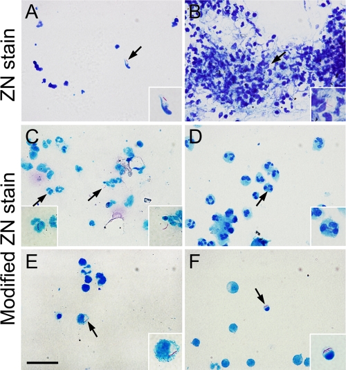
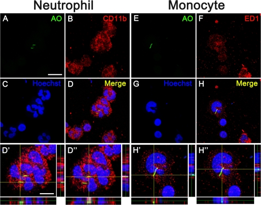
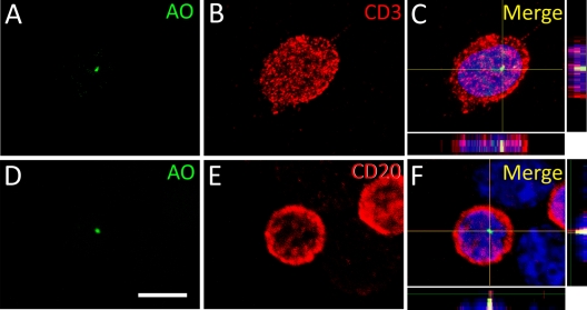
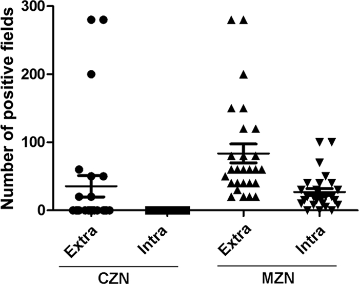
Tuberculous meningitis leads to a devastating outcome, and early diagnosis and rapid chemotherapy are vital to reduce morbidity and mortality. Since Mycobacterium tuberculosis is a kind of cytozoic pathogen and its numbers are very few in cerebrospinal fluid, detecting M. tuberculosis in cerebrospinal fluid from tuberculous meningitis patients is still a challenge for clinicians. Ziehl-Neelsen stain, the current feasible microbiological method for the diagnosis of tuberculosis, often needs a large amount of cerebrospinal fluid specimen but shows a low detection rate of M. tuberculosis. Here, we developed a modified Ziehl-Neelsen stain, involving cytospin slides with Triton processing, in which only 0.5 ml of cerebrospinal fluid specimens was required. This method not only improved the detection rate of extracellular M. tuberculosis significantly but also identified intracellular M. tuberculosis in the neutrophils, monocytes, and lymphocytes clearly. Thus, our modified method is more effective and sensitive than the conventional Ziehl-Neelsen stain, providing clinicians a convenient yet powerful tool for rapidly diagnosing tuberculous meningitis.


Figures

Fig 1
Fig 1
Comparison of the conventional and modified Ziehl-Neelsen (ZN) stain for CSF samples from tuberculous meningitis patients. (A and B) The conventional stain shows damaged cellular structure (A) and cell aggregations (B). No intracellular acid-fast bacilli (AFB) are observed with the conventional method. (C) The modified method can concentrate AFB in the CSF. Intracellular AFB are frequently observed in neutrophils (D), monocytes (E), and lymphocytes (F). Arrows show acid-fast-dye-positive AFB. Insets show higher magnification views of AFB or cells indicated by arrows. Scale bars, 20 (A to F) and 5 μm (insets).
Fig 2
Fig 2
Intracellular distribution of AFB in neutrophils and monocytes on the modified Ziehl-Neelsen stain. Double labeling of AO (A and E, green) with CD11b (B, red) and ED1 (F, red) shows the intracellular location of AFB in neutrophils (A to D) and monocytes (E to H). AO, CD11b, and ED1 label AFB, neutrophils, and monocytes, respectively. The nuclei are stained by Hoechst 33342 (blue). Panels D′, D″, H′, and H″ show higher magnification views of panels D and H in z axis projections. Scale bars, 20 (A to H) and 5 μm (D′, D″, H′, and H″).
Fig 3
Fig 3
Intracellular distribution of AFB in lymphocytes on the modified Ziehl-Neelsen stain. Double labeling of AO (A and D, green) with CD3 (B, red) and CD20 (E, red) shows the intracellular location of AFB in lymphocytes (C and F). The nuclei are stained by Hoechst 33342 (blue). Scale bar, 5 μm.
Fig 4
Fig 4
Comparison of AFB-positive fields by the conventional (CZN) and modified (MZN) Ziehl-Neelsen stain. Three hundred fields on each slide from 48 CSF specimens were observed. Compared to the ZN stain, which reveals no intracellular AFB-positive fields, the modified ZN stain definitely identifies AFB within the immune cells. Moreover, the modified stain reveals more extracellular AFB-positive fields than the conventional ZN stain.

References

    1. Alemán M, GarcíA A, Saab MA. 2002. Mycobacterium tuberculosis-induced activation accelerates apoptosis in peripheral blood neutrophils from patients with active tuberculosis. Am. J. Respir. Cell Mol. Biol. 27:583–592 - PubMed
    1. Alemán M, et al. 2004. Mycobacterium tuberculosis triggers apoptosis in peripheral neutrophils involving toll-like receptor 2 and p38 mitogen protein kinase in tuberculosis patients. Infect. Immun. 72:5150–5158 - PMC - PubMed
    1. Appelberg R, Silva MT. 1989. T cell-dependent chronic neutrophilia during mycobacterial infections. Clin. Exp. Immunol. 78:478–483 - PMC - PubMed
    1. Attallah AM, et al. 2003. Rapid and simple detection of a Mycobacterium tuberculosis circulating antigen in serum using dot-ELISA for field diagnosis of pulmonary tuberculosis. J. Immunoassay Immunochem. 24:73–87 - PubMed
    1. Dora JM, et al. 2008. Polymerase chain reaction as a useful and simple tool for rapid diagnosis of tuberculous meningitis in a Brazilian tertiary care hospital. Braz. J. Infect. Dis. 12:245–247 - PubMed

Publication types

MeSH terms

Substances

LinkOut - more resources