. 2012;7(1):e29920.
doi: 10.1371/journal.pone.0029920. Epub 2012 Jan 6.

Trihydrophobin 1 phosphorylation by c-Src regulates MAPK/ERK signaling and cell migration

Affiliations

Trihydrophobin 1 phosphorylation by c-Src regulates MAPK/ERK signaling and cell migration

Weibin Wu et al. PLoS One. 2012.

Abstract

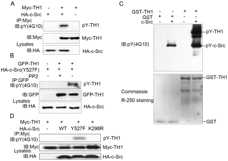
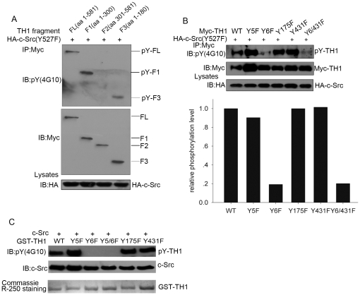
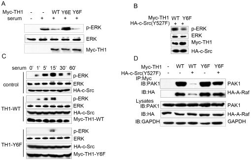
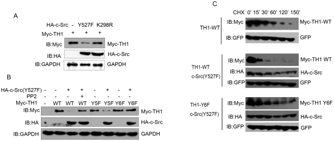
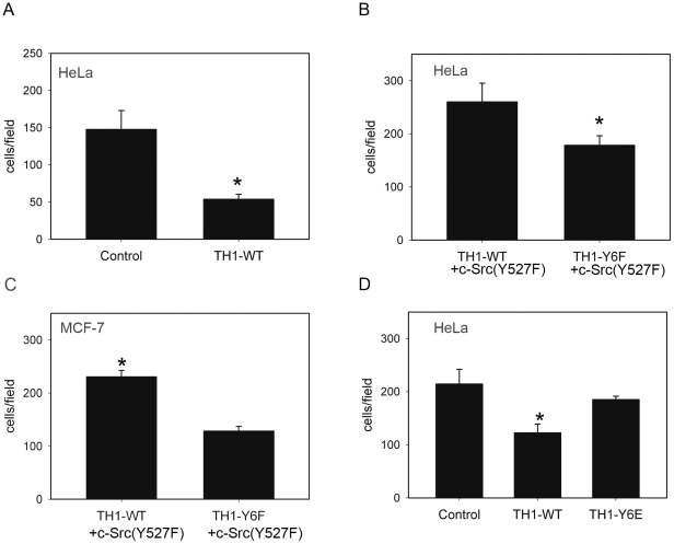
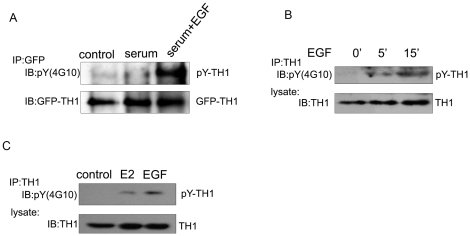
c-Src activates Ras-MAPK/ERK signaling pathway and regulates cell migration, while trihydrophobin 1 (TH1) inhibits MAPK/ERK activation and cell migration through interaction with A-Raf and PAK1 and inhibiting their kinase activities. Here we show that c-Src interacts with TH1 by GST-pull down assay, coimmunoprecipitation and confocal microscopy assay. The interaction leads to phosphorylation of TH1 at Tyr-6 in vivo and in vitro. Phosphorylation of TH1 decreases its association with A-Raf and PAK1. Further study reveals that Tyr-6 phosphorylation of TH1 reduces its inhibition on MAPK/ERK signaling, enhances c-Src mediated cell migration. Moreover, induced tyrosine phosphorylation of TH1 has been found by EGF and estrogen treatments. Taken together, our findings demonstrate a novel mechanism for the comprehensive regulation of Ras/Raf/MEK/ERK signaling and cell migration involving tyrosine phosphorylation of TH1 by c-Src.


Conflict of interest statement

Competing Interests: The authors have declared that no competing interests exist.

Figures

Figure 1
Figure 1. Interaction of c-Src with TH1.
(A) c-Src interacts with TH1. HEK293T cells were transiently transfected with GFP-TH1 and/or HA-c-Src as described under “Experimental Procedures”. At 36 h after transfection, the cells were lysed, immunoprecipitated (IP) with 2 µg of anti-GFP antibody, immunoblotted (IB) with an anti-HA antibody. (B) GST-pull down assay detects interaction of c-Src with TH1 in vitro. Recombinant GST-TH1 or GST was incubated with HeLa cell lysates, precipitated by glutathione-Sepharose beads. The proteins were subjected to western blot and probed with anti-c-Src and anti-GST antibodies. (C) Interaction of endogenous c-Src with TH1 in HeLa cells. HeLa cells were lysed, immunoprecipitated with rabbit anti-TH1 antibody or control IgG. The proteins were subjected to western blot and probed with anti-c-Src antibody. (D) Colocalization of c-Src with TH1. HeLa cells were stained with anti-TH1 and anti-c-Src antibodies together with FITC-conjugated and Rhodamine-conjugated secondary antibodies, observed under a confocal fluorescence microscope. FITC-TH1 shown in green, Rhodamine-c-Src shown in red, the overlay image shown in yellow, Scale bar: 10 µm. (E) Interaction of TH1 with c-Src mutant under serum deprivation condition. The 293T cells transfected with GFP-TH1 and HA-c-Src mutant were serum starved or stimulated as indicated, immunoprecipitated with anti-GFP antibody, followed by western blot with anti-HA antibody. All the data are representative of at least three independent experiments.
Figure 2
Figure 2. Phosphorylation of TH1 by c-Src.
(A) c-Src phosphorylates TH1. COS-7 cells transfected as indicated were lysed, precipitated with anti-Myc antibody bound agarose beads, followed by western blot with anti-phosphotyrosine (4G10) and anti-Myc antibodies. (B) Tyrosine phosphorylation of TH1 by c-Src is abolished by PP2, a specific inhibitor of c-Src. HEK293T cells transfected with GFP-TH1 and HA-c-Src(Y527F) were treated with 10 µM PP2 for 30 min and then immunoprecipitated with anti-GFP antibody, followed by western blot with anti-phosphotyrosine (4G10) and anti-GFP antibodies. (C) c-Src phosphorylates TH1 in vitro. Recombinant GST-TH1 or GST was incubated with recombinant c-Src (5 U) in a kinase buffer as described under “Experimental Procedures”. The reaction mixtures were subjected to western blot and probed with anti-pY (4G10) antibody (upper panel) and then the PVDF membrane was subjected to Coomassie brilliant blue R-250 staining (lower panel). (D) Phosphorylation of TH1 by c-Src mutant under serum deprivation condition. COS-7 cells were transfected with Myc-tagged TH1 and HA-c-Src mutant as indicated, serum-starved for 18 h, lysed and precipitated with anti-Myc antibody bound agarose beads, followed by western blot with anti-phosphotyrosine (4G10) and anti-Myc antibodies. All the data are representative of at least three independent experiments.
Figure 3
Figure 3. c-Src phosphorylates TH1 at Tyr-6.
(A) Phosphorylation of TH1 fragments by c-Src. COS-7 cells were transfected with HA-c-Src (Y527F) and constructs expressing the indicated Myc-tagged TH1 fragments, immunoprecipitated with anti-Myc antibody, followed by western blot with anti-phosphotyrosine and anti-Myc antibodies. (B) Tyr-6 is the major phosphorylation site for TH1. COS-7 cells were transfected with HA-c-Src (Y527F) and constructs expressing the indicated Myc-tagged TH1 mutants, immunoprecipitated with anti-Myc antibody, followed by western blot with anti-phosphotyrosine and anti-Myc antibodies. The quantification of immunoblot intensity is shown at the bottom panel. (C) c-Src phosphorylates TH1 at Tyr-6 in vitro. Recombinant c-Src was incubated with GST-TH1 or GST-TH1 mutant as indicated at 30°C for 30 min and then subjected to western blot and probed with anti-phosphotyrosine and anti-c-Src antibodies. Coomassie brilliant blue staining showed the input of GST-TH1 mutant proteins. All the data are representative of at least three independent experiments.
Figure 4
Figure 4. Tyr-6 phosphorylation of TH1 impairs its inhibition on MAPK/ERK signaling.
(A) TH1-Y6E mutant lost its inhibition on ERK phosphorylation. HeLa cells were transfected either with Myc-TH1-WT (wild type of TH1), with Myc-TH1-Y6E (the constitutive phosphorylation form of TH1), or with TH1-Y6F (the dephosphorylation form of TH1) and then serum starved for 18 h, stimulated with or without 20% serum for 15 min, lysed, followed by western blot with antibodies against phospho-ERK, total ERK and Myc. (B) TH1-Y6F mutant shows increased inhibition on ERK phosphorylation. HeLa cells transfected with Myc-TH1-WT or Myc-TH1-Y6F were cotransfected with HA-c-Src (Y527F), lysed, followed by western blot with antibodies against phospho-ERK, total ERK, Myc and HA. (C) Time course of ERK phosphorylation. HeLa cells transfected either with Myc-TH1-WT, or with Myc-TH1-Y6F were cotransfected with HA-c-Src, serum starved for 18 h, stimulated with 20% serum for the indicated times. Cell lysates were subjected to western blot with antibodies against phospho-ERK, total ERK, Myc and HA. (D) Phosphorylation of TH1 by c-Src decreases its interaction with PAK1 and A-Raf. HeLa cells transiently transfected with HA-A-Raf and Myc-TH1-WT or Myc-TH1-Y6F were cotransfected with c-Src (Y527F) or control vector. At 36 h after transfection, cell lysates were immunoprecipitated with anti-Myc antibodies. Myc–TH1 associated HA-A-Raf or PAK1 was monitored by anti-HA or anti-PAK1 antibody in the following western blot analyses. Whole cell lysates were also subjected to western blot with anti-HA and anti-PAK1 antibodies. All the data are representative of at least three independent experiments.
Figure 5
Figure 5. Tyr-6 phosphorylation of TH1 impacts its stability in HeLa cells.
(A) c-Src (Y527F) lowers TH1 expression level. HeLa cells transiently transfected either with HA-c-Src (Y527F), with HA-c-Src (K298R), or with control vector were cotransfected with Myc-TH1 for 36 h, pretreated with 100 µM pervanadate, lysed, subject to western blot with antibodies against Myc, HA and GAPDH as internal control. (B) Both PP2 treatment and TH1-Y6F mutant inhibit c-Src mediated TH1 lowering. HeLa cells transfected with Myc-tagged TH1 mutant and HA-c-Src(Y527F) or control vector as indicated for 36 h were treated with 10 µM PP2 or vehicle for 1 h, pretreated with 100 µM pervanadate, lysed, subjected to western blot with antibodies against Myc, HA and GAPDH as internal control. (C) Time course of TH1 degradation. HeLa cells transfected with Myc-TH1-WT or Myc-TH1-Y6F and GFP expression vector (GFP level was used as transfection control) were cotransfected with HA-c-Src (Y527F) or control vector as indicated for 36 h, treated with 50 µg/ml cycloheximide (CHX) to inhibit protein synthesis, and 100 µM pervanadate to inhibit dephosphorylation. Lysates were harvested at the time points as indicated, subjected to western blot with antibodies against Myc and HA.
Figure 6
Figure 6. Enhanced cell migration through Tyr-6 phosphorylation of TH1.
(A) TH1-WT inhibits HeLa cell migration. HeLa cells were transfected with Myc-TH1-WT or control vector. Transwell cell migration assay was performed as described under “Experimental Procedures” (n = 4, mean ± SD, P<0.01 vs. Control). Statistical significance was determined by the Student's t test. (B–C) TH1-Y6F mutant shows increased inhibition on HeLa and MCF-7 cells migration. HeLa and MCF-7 cells transfected with Myc-TH1-WT or Myc-TH1-Y6F were cotransfected with HA-c-Src (Y527F) and then measured migration by Transwell cell migration assay (n = 3, mean ± SD, P<0.05 TH1-WT vs. TH1-Y6F). (D) Inhibition of cell migration is abolished in TH1-Y6E mutant. HeLa cells were transfected with Myc-TH1-WT, Myc-TH1-Y6E or control vector, and then measured migration by Transwell cell migration assay (n = 4, mean ± SD, P<0.01 TH1-WT vs. Control, P = 0.2 TH1-Y6F vs. Control).
Figure 7
Figure 7. Induced Tyrosine Phosphorylation of TH1 by EGF and Estrogen Stimulations.
(A) Induced tyrosine phosphorylation of TH1 by EGF in HEK293T cells. HEK293T cells over-expressing GFP-TH1 were serum starved for 18 h, pretreated with 50 µM pervanadate for 15 min and incubated with or without 100 ng/ml EGF for 15 min in the presence of 10% fetal bovine serum. Cell lysates were immunoprecipitated with antibody against GFP. The tyrosine phosphorylation of TH1 was analyzed with pY (4G10). The same blot was striped and re-probed with anti-GFP antibody. (B) Induced tyrosine phosphorylation of TH1 by EGF in A549 cells. A549 cells were serum starved for 18 h, pretreated with 100 µM pervanadate for 15 min, stimulated with 100 ng/ml EGF for 5 min and 15 min in the presence of 10% fetal bovine serum. Cell lysates were immunoprecipitated with TH1 antibody. The tyrosine phosphorylation of TH1 was analyzed with pY (4G10). (C) Induced tyrosine phosphorylation of TH1 by Estrogen and EGF in MCF-7 cells. MCF-7 cells were serum and estrogen deprived, pretreated with 100 µM pervanadate for 15 min, and stimulated with10 nM 17β-estradiol (E2) for 2 min or 100 ng/ml EGF for 15 min in the presence of 10% fetal bovine serum. Tyrosine phosphorylation of TH1 was analyzed as in (B).

Similar articles

Cited by

References

    1. Yeatman TJ. A renaissance for SRC. Nat Rev Cancer. 2004;4:470–480. - PubMed
    1. Frame MC. Src in cancer: deregulation and consequences for cell behaviour. Biochim Biophys Acta. 2002;1602:114–130. - PubMed
    1. Soriano P, Montgomery C, Geske R, Bradley A. Targeted disruption of the c-src proto-oncogene leads to osteopetrosis in mice. Cell. 1991;64:693–702. - PubMed
    1. Ishizawar R, Parsons SJ. c-Src and cooperating partners in human cancer. Cancer Cell. 2004;6:209–214. - PubMed
    1. Mayer EL, Krop IE. Advances in targeting SRC in the treatment of breast cancer and other solid malignancies. Clin Cancer Res. 2010;16:3526–3532. - PubMed

Publication types