. 2012 Jan 13;3(1):2.
doi: 10.1186/2041-9139-3-2.

Lim homeobox genes in the Ctenophore Mnemiopsis leidyi: the evolution of neural cell type specification

Affiliations

Lim homeobox genes in the Ctenophore Mnemiopsis leidyi: the evolution of neural cell type specification

David K Simmons et al. Evodevo. .

Abstract

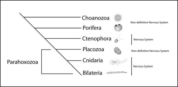
Background: Nervous systems are thought to be important to the evolutionary success and diversification of metazoans, yet little is known about the origin of simple nervous systems at the base of the animal tree. Recent data suggest that ctenophores, a group of macroscopic pelagic marine invertebrates, are the most ancient group of animals that possess a definitive nervous system consisting of a distributed nerve net and an apical statocyst. This study reports on details of the evolution of the neural cell type specifying transcription factor family of LIM homeobox containing genes (Lhx), which have highly conserved functions in neural specification in bilaterian animals.

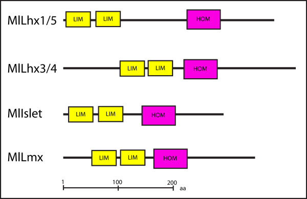
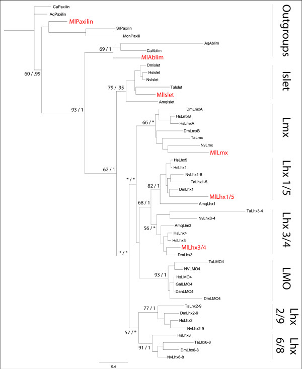
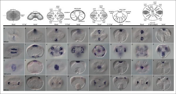
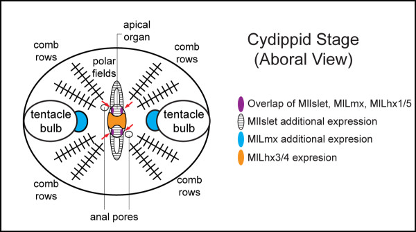
Results: Using next generation sequencing, the first draft of the genome of the ctenophore Mnemiopsis leidyi has been generated. The Lhx genes in all animals are represented by seven subfamilies (Lhx1/5, Lhx3/4, Lmx, Islet, Lhx2/9, Lhx6/8, and LMO) of which four were found to be represented in the ctenophore lineage (Lhx1/5, Lhx3/4, Lmx, and Islet). Interestingly, the ctenophore Lhx gene complement is more similar to the sponge complement (sponges do not possess neurons) than to either the cnidarian-bilaterian or placozoan Lhx complements. Using whole mount in situ hybridization, the Lhx gene expression patterns were examined and found to be expressed around the blastopore and in cells that give rise to the apical organ and putative neural sensory cells.

Conclusion: This research gives us a first look at neural cell type specification in the ctenophore M. leidyi. Within M. leidyi, Lhx genes are expressed in overlapping domains within proposed neural cellular and sensory cell territories. These data suggest that Lhx genes likely played a conserved role in the patterning of sensory cells in the ancestor of sponges and ctenophores, and may provide a link to the expression of Lhx orthologs in sponge larval photoreceptive cells. Lhx genes were later co-opted into patterning more diversified complements of neural and non-neural cell types in later evolving animals.


Figures

Figure 1
Figure 1
Phylogenetic relationships of 'basal metazoa'. Phylogenetic relationships of early branching metazoan taxa, based on data from recent studies [45-47]. Non-definitive nervous systems refer to the lack of morphological and physiological data to confirm their existence.
Figure 2
Figure 2
Domain structure of Mnemiopsis Lhx genes. The domain structure of the Mnemiopsis leidyi Lhx genomic complement was predicted by using the SMART database [48]. Yellow boxes indicate the tandem LIM zinc finger binding domains, pink boxes indicate the homeodomain sequence.
Figure 3
Figure 3
Alignment of Lmx homeodomain sequence. Alignment of the Lmx genes homeodomain region, showing the conservation of two intervening introns labeled (0). Mnemiopsis's first intron position is 3' to the highly conserved position in other taxa. The second intron position is shared with other taxa, but not with the anthozoan Nematostella. Dm,Drosophila Melanogaster; Hs,Homo sapiens; Ml, Mnemiopsis leidyi; Nv,Nematostella vectensis; Ta,Trichoplax adhaerens.
Figure 4
Figure 4
Phylogenetic Tree of Lhx genes. Maximum likelihood and Bayesian analysis consensus tree based on the tandem LIM domains and homeodomain, support values are indicated by Likelihood > 50/Bayesian analyses >.95, low support indicated by an asterisk. Construction of the Lhx gene orthology was conducted in RaxML v7.0.0 [82] using maximum likelihood analysis under the JTT I+G model of evolution determined by ProtTest v1.4 analysis [83]. A total of 1,000 searches was performed and 500 bootstrap replicates were applied to the tree with the best likelihood score used to generate branch support values. Bayesian analysis used two runs of Mr Bayes after 2,000,000 generations, JTT I+G, burnin = 5000 trees. Tree was rooted to the unicellular choanoflagellate Monosiga brevicollis LIM domain containing gene paxilin. Aq, Amphimedon queenslandica; Ca, Capsaspora owczarzaki; Dan, Danio rerio; Dm, Drosophila melanogaster; Gal, Gallus gallus; Hs, Homo sapiens;, Ml, Mnemiopsis leidyi; Mon, Monosiga brevicollis; Nv, Nematostella vectensis; Ta,Trichoplax adhaerens.
Figure 5
Figure 5
Whole mount in situ hybridization of Lhx genes in Mnemiopsis. Whole mount in situ hybridization of MlIslet (A-H), MlLhx1/5 (I-P), MlLhx3/4 (Q-X), and MlLmx (Y-F'), purple staining represents the localization of detected mRNA transcripts. The above cartoons are representations of the underlying corresponding stages of development. All views are either aboral or lateral where indicated, with the asterisk representing the blastopore and future mouth. (A-H) MlIslet is expressed in aboral ectodermal cells, adjacent to MlLhx3/4, that give rise to part of the apical organ floor as well as the polar fields. (I-P) MlLhx1/5 is expressed around the blastopore, which gives rise to the pharynx. It is also expressed in mesodermal cells that give rise to the photocytes, which underlie four of the comb rows. There is late expression in four small groups of cells in the apical organ which overlap with cells that give rise to proposed photosensory cells. (Q-X) MlLhx3/4 is expressed in the aboral ectoderm, in a large group of cells that gives rise to part of the apical organ. (Y-F') MlLmx is expressed in two groups of cells that give rise to part of the tentacle bulb apparatus. Expression is also found in the same four groups of cells in the apical organ that overlap with the MlLhx1/5 expression.
Figure 6
Figure 6
Lhx expression diagram. Summary diagram of the aboral overlapping and non-overlapping Lhx expression domains during the cydippid stage of Mnemiopsis. Overlapping expression is found in the four groups of cells within the apical organ that correspond to putative photosensory cells, indicated by red arrows. Non-overlapping domains include portions of the apical organ and associated polar fields, highly innervated sensory and nervous structures.

Similar articles

Cited by

References

    1. Way JC, Chalfie M. mec-3, a homeobox-containing gene that specifies differentiation of the touch receptor neurons in C. elegans. Cell. 1988;54:5–16. doi: 10.1016/0092-8674(88)90174-2. - DOI - PubMed
    1. Freyd G, Kim SK, Horvitz HR. Novel cysteine-rich motif and homeodomain in the product of the Caenorhabditis elegans cell lineage gene lin-11. Nature. 1990;344:876–879. doi: 10.1038/344876a0. - DOI - PubMed
    1. Karlsson O, Thor S, Norberg T, Ohlsson H, Edlund T. Insulin gene enhancer binding protein Isl-1 is a member of a novel class of proteins containing both a homeo- and a Cys-His domain. Nature. 1990;344:879–882. doi: 10.1038/344879a0. - DOI - PubMed
    1. Hobert O, Westphal H. Functions of LIM-homeobox genes. Trends Genet. 2000;16:75–83. doi: 10.1016/S0168-9525(99)01883-1. - DOI - PubMed
    1. Shirasaki R, Pfaff SL. Transcriptional codes and the control of neuronal identity. Annu Rev Neurosci. 2002;25:251–281. doi: 10.1146/annurev.neuro.25.112701.142916. - DOI - PubMed