Review
. 2012 Feb;23(1):149-72, xii.
doi: 10.1016/j.pmr.2011.11.014.

The paradox of muscle hypertrophy in muscular dystrophy

Affiliations
Review

The paradox of muscle hypertrophy in muscular dystrophy

Joe N Kornegay et al. Phys Med Rehabil Clin N Am. 2012 Feb.

Abstract

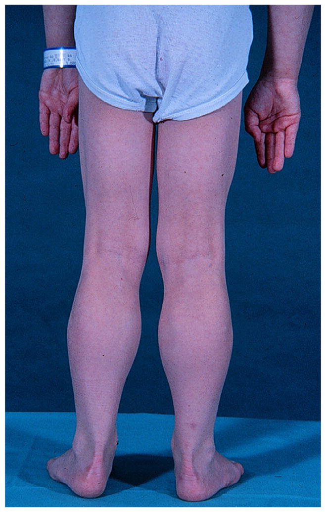
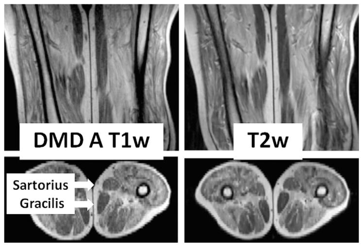
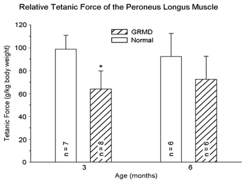
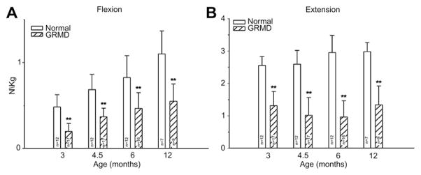
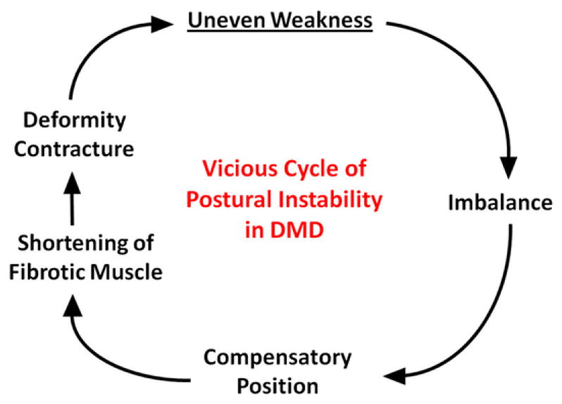
Mutations in the dystrophin gene cause Duchenne and Becker muscular dystrophy in humans and syndromes in mice, dogs, and cats. Affected humans and dogs have progressive disease that leads primarily to muscle atrophy. Mdx mice progress through an initial phase of muscle hypertrophy followed by atrophy. Cats have persistent muscle hypertrophy. Hypertrophy in humans has been attributed to deposition of fat and connective tissue (pseudohypertrophy). Increased muscle mass (true hypertrophy) has been documented in animal models. Muscle hypertrophy can exaggerate postural instability and joint contractures. Deleterious consequences of muscle hypertrophy should be considered when developing treatments for muscular dystrophy.


Figures

Fig. 1
Fig. 1
Nine-year-old boy with DMD demonstrating characteristic calf hypertrophy. (Courtesy of James F. Howard Jr.)
Fig. 2
Fig. 2
T1- and T2-weighted magnetic resonance images of the proximal leg of a boy with DMD in the anteroposterior (top) and transverse (bottom) planes. The transverse image is at the level of the midfemur. Most muscles have been partially to near totally replaced with signal-intense material compatible with fat. Note that the subcutaneous fat has comparable signal intensity in both T1- and T2-weighted images. In contrast to the properties of most muscles seen, the sartorius and gracilis muscles (arrows) are largely unaffected.
Fig. 3
Fig. 3
Tetanic force corrected for body weight (g/kg) generated by the peroneus longus muscle of normal and GRMD dogs at 3 and 6 months of age. Values for the GRMD dogs are lower (P<.05)* at 3 months. However, although body-weight-corrected force is proportionally lower in normal dogs at 6 months, it has increased in GRMD dogs, such that values for the 2 groups are no longer significantly different. (Data from Kornegay JN, Sharp NJ, Bogan DJ, et al. Contraction tension and kinetics of the peroneus longus muscle in golden retriever muscular dystrophy. J Neurol Sci 1994;123:100–7.)
Fig. 4
Fig. 4
Tetanic force, corrected for body weight (N/kg), generated by tarsal joint flexors (left) and extensors (right) from normal dogs and GRMD dogs at 3, 4.5, 6, and 12 months of age. Values for GRMD dogs are lower (P<.01 for all) than those of normal dogs at all ages. However, the differential between GRMD and normal dogs differs. Flexion values are especially low at 3 months, whereas extension is affected more at later ages. (From Kornegay JN, Bogan DJ, Bogan JR, et al. Contraction force generated by tibiotarsal joint flexion and extension in dogs with golden retriever muscular dystrophy. J Neurol Sci 1999;166:119; with permission.)
Fig. 5
Fig. 5
Vicious cycle of postural instability that leads to loss of ambulation in patients with DMD. Uneven weakness leads to imbalance and compensatory postural changes that ultimately result in shortening of muscles and contractures. The process is self-perpetuating. (Modified from Roy L, Gibson DA. Pseudohypertrophic muscular dystrophy and its surgical management: review of 30 patients. Can J Surg 1970;13:14; with permission.)
Fig. 6
Fig. 6
Lateral (A) and ventrodorsal (B) radiographs of the pelvis of a 4-year-old GRMD dog and a normal dog (C and D) for comparison. Note the marked vertical tilt of the pelvis in the GRMD dog (A), so that the angle formed by the wing of the ilium and lumbar spine is much more acute at approximately 90° (angle marked by lines within the red circle). The wings of the ilia flare laterally in the GRMD dog (red circle in B). (From Brumitt JW, Essman SC, Kornegay JN, et al. Radiographic features of Golden Retriever muscular dystrophy. Vet Radiol Ultrasound 2006;47:578; with permission.)
Fig. 7
Fig. 7
T2-weighted magnetic resonance images with fat signal suppression (FS) of pelvic limb muscles at midthigh from 3 crossbred GRMD and myostatin-heterozygous (Mstn+/) dogs from the first litter. Note the proportional enlargement of the sartorius and hamstring muscles and the associated atrophy/hypoplasia of the quadriceps of the dystrophic Mstn+/+ dog, Flash, relative to the normal dog, Racer, and the even more dramatic differential size of these muscles in the dystrophic Mstn+/ dog, Dash (also see volumetric measurements from these sections in Table 2). Segmentation was done using ITK-SNAP (http://www.itksnap.org/pmwiki/pmwiki.php).

Similar articles

Cited by

References

    1. Tyler KL. Origins and early descriptions of “Duchenne muscular dystrophy”. Muscle Nerve. 2003;28:402–22. - PubMed
    1. Hoffman EP, Brown RH, Kunkel LM. Dystrophin: the protein product of the Duchenne muscular dystrophy locus. Cell. 1998;51:919–28. - PubMed
    1. Prior TW, Bridgeman SJ. Experience and strategy for the molecular testing of Duchenne muscular dystrophy. J Mol Diagn. 2005;7:317–26. - PMC - PubMed
    1. Malhotra S, Hart K, Klamut H, et al. Frame-shift deletions in patients with Duchenne and Becker muscular dystrophy. Science. 1988;242:755–9. - PubMed
    1. Koenig M, Beggs AH, Moyer M, et al. The molecular basis for Duchenne versus Becker muscular dystrophy: correlation of severity with type of deletion. Am J Hum Genet. 1989;45:498–506. - PMC - PubMed

MeSH terms