. 2012 Jan 12:11:4.
doi: 10.1186/1476-4598-11-4.

Enhancement of lung tumorigenesis in a Gprc5a Knockout mouse by chronic extrinsic airway inflammation

Affiliations

Enhancement of lung tumorigenesis in a Gprc5a Knockout mouse by chronic extrinsic airway inflammation

Peter Barta et al. Mol Cancer. .

Abstract

Background: Although cigarette smoking is the principal cause of lung carcinogenesis, chronic obstructive pulmonary disease (COPD), an inflammatory disease of the lung, has been identified as an independent risk factor for lung cancer. Bacterial colonization, particularly with non-typeable Haemophilus influenzae (NTHi), has been implicated as a cause of airway inflammation in COPD besides cigarette smoke. Accordingly, we hypothesized that lung cancer promotion may occur in a chronic inflammatory environment in the absence of concurrent carcinogen exposure.

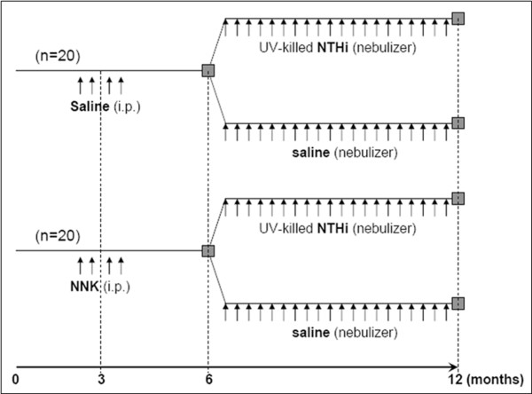
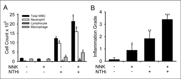
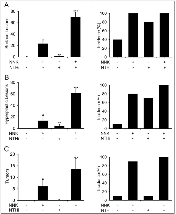
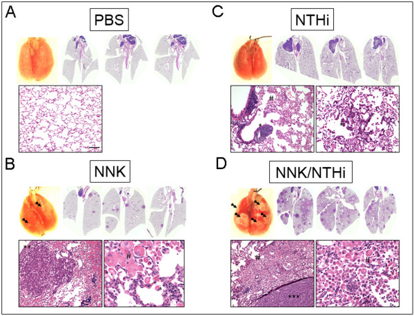
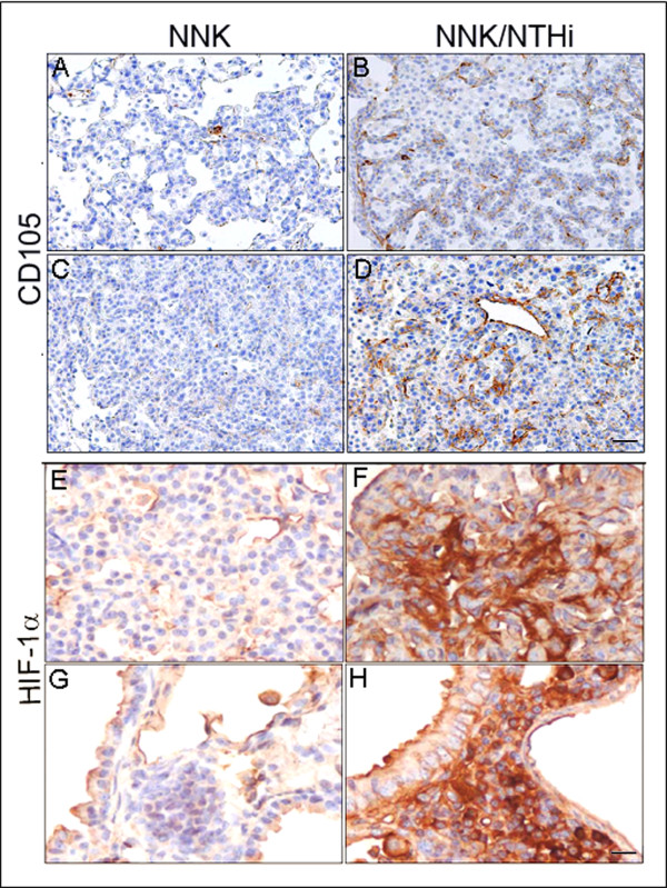
Results: Herein, we investigated the effects of bacterial-induced COPD-like inflammation and tobacco carcinogen-enhanced tumorigenesis/inflammation in the retinoic acid inducible G protein coupled receptor knock out mouse model (Gprc5a-/- mouse) characterized by late-onset, low multiplicity tumor formation. Three-month-old Gprc5a-/- mice received 4 intraperitoneal injections of the tobacco-specific carcinogen, NNK, followed by weekly exposure to aerosolized NTHi lysate for 6 months. The numbers of inflammatory cells in the lungs and levels of several inflammatory mediators were increased in Gprc5a-/- mice treated with NTHi alone, and even more so in mice pretreated with NNK followed by NTHi. The incidence of spontaneous lung lesions in the Gprc5a-/- mice was low, but NTHi exposure led to enhanced development of hyperplastic lesions. Gprc5a-/- mice exposed to NNK alone developed multiple lung tumors, while NTHi exposure increased the number of hyperplastic foci 6-fold and the tumor multiplicity 2-fold. This was associated with increased microvessel density and HIF-1α expression.

Conclusion: We conclude that chronic extrinsic lung inflammation induced by bacteria alone or in combination with NNK enhances lung tumorigenesis in Gprc5a-/- mice.


Figures

Figure 1
Figure 1
Experimental design. Gprc5a-/- mice were randomized to 4 consecutive, weekly injections of NNK or saline between the ages of 10 and 14 weeks, followed by exposure to an aerosolized UV-killed lysate of NTHi or saline between the ages of 6 and 12 months.
Figure 2
Figure 2
Analysis of lung inflammation. (A) Total and lineage-specific leukocyte numbers in bronchoalveolar lavage fluid (BALF) one day after the last NTHi or saline aerosol exposure are shown (n = 4, mean ± SE, *= P < 0.05 for NTHi or NNK/NTHi-exposed Gprc5a-/- mice vs PBS or NNK-exposed Gprc5a-/- mice). (B) Histologic grading of inflammation in lung tissue from each treatment group (n = 6, mean ± SE, *= P < 0.05 for NNK vs PBS, **= P < 0.05 for NTHi vs NNK, and ***= P < 0.05 for NNK/NTHi-exposed Gprc5a-/- mice vs NTHi-exposed Gprc5a-/- mice).
Figure 3
Figure 3
Analysis of lung carcinogenesis. (A) Lung surface lesion numbers (left) and incidence of lesion development (right) in Gprc5a-/- mice. (B) Number and incidence of hyperplastic lesions in the lungs of Gprc5a-/- mice. (C) Number and incidence of tumoral lesions in the lungs of Gprc5a-/- mice. (Incidence is calculated as the percentage of mice with surface lesions, hyperplastic lesions, or tumoral lesions among the total mice in the same study group, and presented as mean ± SE, *= P < 0.05 for NNK vs PBS, **= P < 0.05 for NTHi vs NNK, and ***= P < 0.05 for NNK/NTHi-exposed Gprc5a-/- mice vs NTHi-exposed Gprc5a-/- mice).
Figure 4
Figure 4
Gross and microscopic analysis of lungs in Gprc5a-/- mice. (A) Surface of freshly removed, formalin-filled control lungs of 12 months old Gprc5a-/- mice lacks visible lesions. Scans of H&E stained lung sections (2X) and microscopic image (10X) shows no lesion. (B) A few surface lesions (double arrows) were seen after NNK treatment. Scans of H&E stained cross sections of NNK-treated lung (2X) and high magnification (10X) microscopic images demonstrate the appearance of several papillary adenomas. (C) NTHi treatment led to perivascular and peribronchiolar lymphocyte infiltration (**) and characteristic alveolar accumulation of acidophilic macrophages, often resulting in confluent areas of acidophil macrophage pneumonia (AMP, #). AMP strongly co-localized with the widespread appearance of NTHi-induced premalignant, hyperplastic lesions (10X). (D) NNK+NTHi combined treatment led to confluent lesions on the lung surface (double arrows). A high number of pulmonary adenomas and areas of AMP are seen on the H&E scans (2X). Pulmonary hyperplasia with co-localized acidophilic macrophages (#) and papillary adenocarcinomas (***) with surrounding of heavy inflammatory infiltration characterized by the accumulation of macrophages and lymphocytes are seen on the microscopic images (10X, scale bar = 100 μm).
Figure 5
Figure 5
Analysis of microvascular density and HIF-1α activity. Microvascular density of pulmonary tumors was significantly higher in hyperplastic (B) and tumoral lesions (D) of NNK/NTHi treated mice compared to the NNK treated mice (A, and C) detected by CD105 immunostaining (10X, scale bar = 100 μm). HIF-1α immunostaining after NNK/NTHi combined treatment showed hot-spots of high stromal expression in tumors (F), and high expression in perivascular-peribronchiolar lymphocytes (H). In contrast, low, homogenous expression of HIF-1α was detected in the tumors (E) and perivascular-peribronchiolar lymphocytes (G) of NNK treated mice (40X, scale bar = 25 μm).

References

    1. Jemal A, Siegel R, Ward E, Hao Y, Xu J, Thun MJ. Cancer statistics. CA Cancer J Clin. 2009;59:225–249. doi: 10.3322/caac.20006. - DOI - PubMed
    1. Stellman SD, Takezaki T, Wang L, Chen Y, Citron ML, Djordjevic MV, Harlap S, Muscat JE, Neugut AI, Wynder EL. et al.Smoking and lung cancer risk in American and Japanese men: an international case-control study. Cancer Epidemiol Biomarkers Prev. 2001;10:1193–1199. - PubMed
    1. Schottenfield D. In: Lung cancer: Principles and Practice. Pass HIMJBJDHTATMJD, editor. Philadelphia: Lippincott, Williams and Wilkins; 2000. Etiology and epidemiology of lung cancer; pp. 367–388.
    1. Mannino DM, Aguayo SM, Petty TL, Redd SC. Low lung function and incident lung cancer in the United States: data From the First National Health and Nutrition Examination Survey follow-up. Arch Intern Med. 2003;163:1475–1480. doi: 10.1001/archinte.163.12.1475. - DOI - PubMed
    1. Turner MC, Chen Y, Krewski D, Calle EE, Thun MJ. Chronic obstructive pulmonary disease is associated with lung cancer mortality in a prospective study of never smokers. Am J Respir Crit Care Med. 2007;176:285–290. doi: 10.1164/rccm.200612-1792OC. - DOI - PubMed

Publication types

MeSH terms

Substances