Review
. 2012 Jun;1818(6):1457-65.
doi: 10.1016/j.bbamem.2011.12.026. Epub 2012 Jan 3.

VDAC structure, selectivity, and dynamics

Affiliations
Review

VDAC structure, selectivity, and dynamics

Marco Colombini. Biochim Biophys Acta. 2012 Jun.

Abstract

VDAC channels exist in the mitochondrial outer membrane of all eukaryotic organisms. Of the different isoforms present in one organism, it seems that one of these is the canonical VDAC whose properties and 3D structure are highly conserved. The fundamental role of these channels is to control the flux of metabolites between the cytosol and mitochondrial spaces. Based on many functional studies, the fundamental structure of the pore wall consists of one α helix and 13 β strands tilted at a 46° angle. This results in a pore with an estimated internal diameter of 2.5nm. This structure has not yet been resolved. The published 3D structure consists of 19 β strands and is different from the functional structure that forms voltage-gated channels. The selectivity of the channel is exquisite, being able to select for ATP over molecules of the same size and charge. Voltage gating involves two separate gating processes. The mechanism involves the translocation of a positively charged portion of the wall of the channel to the membrane surface resulting in a reduction in pore diameter and volume and an inversion in ion selectivity. This mechanism is consistent with experiments probing changes in selectivity, voltage gating, kinetics and energetics. Other published mechanisms are in conflict with experimental results. This article is part of a Special Issue entitled: VDAC structure, function, and regulation of mitochondrial metabolism.


Figures

Fig. 1
Fig. 1
The open state of VDAC mediates ATP flux and VDAC closure stops this flux despite the closed state being conductive and having an estimated pore radius of 0.9 nm. The channels were reconstituted into a planar phospholipid membrane under voltage clamp conditions. The number of open and closed channels was monitored continuously as was the flux of ATP. The presence of a large ATP gradient shifted the voltage dependent characteristics as described[72]. Reprinted from[8].
Fig 2
Fig 2
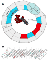
Fig. 2A. A drawing of the top view of the open state of a VDAC channel using the constraints from experimental results[39]. Functional studies show that the pore is composed of 1 α helix and 13 β strands. Structural studies report that the β strands are tilted by 46° [20]. Thus from the top view, the apparent backbone to backbone distances for the β strands is 6.8 nm and the α helix is elongated in one dimension to 14 nm. The resulting pore diameter is 2.5 nm, in good agreement with experimental estimates. The colors on the inner wall refer to the net charge of that region based on the folding pattern shown in “B”. Blue is positive, gray is neutral, and red is negative. The molecule of ATP in the center is scaled to the size of the channel and oriented according to the charge on the channel. 2B. The folding pattern of VDAC for human VDAC1 is based on results from the functional studies[39]. As these do not define the ends of each beta strand, the endings were defined based on the NMR structure[33]. The charged residues were color-coded as above. The dotted lines are the arbitrarily defined regions whose net charge is indicated in “A”.
Fig. 3
Fig. 3
The gating process of VDAC consisting of one open state and two closed states: one of these achieved at positive potentials and the other at negative potentials. The black region is the positively-charged, mobile, voltage-sensor domain. Its movement out of the channel results in a reduced diameter, reduced volume, and inverted ion selectivity. Each closed state is actually a population of closed states.
Fig. 4
Fig. 4
Energetics and voltage-dependent kinetics of the VDAC gating process. The data was obtained from ref [60] and these are the results of experiments performed on VDAC from N. crassa reconstituted into planar membranes made from solvent-free soybean phospholipid monolayers. A. The rate constants are the means ± SE of 5 and 4 experiments at each voltage for the closing and opening rates resp. B. The data in “A” were converted to free energy of activation, as described in the text. The values for the rates of opening at positive and negative potentials was pooled into two points (filled symbols; one for negative and one for positive potentials) because there is no voltage dependence of this rate except for the sign of the potential used. The error bars were included but are almost always smaller than the symbol. The straight line is merely the relationship between the rate constant and the activation energy. For the closing rates, the voltage-dependent energy, nFV, was added to the calculated energy barrier to obtain the square symbols. These square symbols represent the true energy barrier for the conformational change from the open to the closed conformation.
Fig. 4
Fig. 4
Energetics and voltage-dependent kinetics of the VDAC gating process. The data was obtained from ref [60] and these are the results of experiments performed on VDAC from N. crassa reconstituted into planar membranes made from solvent-free soybean phospholipid monolayers. A. The rate constants are the means ± SE of 5 and 4 experiments at each voltage for the closing and opening rates resp. B. The data in “A” were converted to free energy of activation, as described in the text. The values for the rates of opening at positive and negative potentials was pooled into two points (filled symbols; one for negative and one for positive potentials) because there is no voltage dependence of this rate except for the sign of the potential used. The error bars were included but are almost always smaller than the symbol. The straight line is merely the relationship between the rate constant and the activation energy. For the closing rates, the voltage-dependent energy, nFV, was added to the calculated energy barrier to obtain the square symbols. These square symbols represent the true energy barrier for the conformational change from the open to the closed conformation.

Similar articles

Cited by

References

    1. Colombini M. Candidate for the permeability pathway of the outer mitochondrial-membrane. Nature. 1979;279:643–645. - PubMed
    1. Schein SJ, Colombini M, Finkelstein A. Reconstitution in planar lipid bilayers of a voltage-dependent anion-selective channel obtained from Paramecium mitochondria. J Membr Biol. 1976;30:99–120. - PubMed
    1. Colombini M. Voltage gating in the mitochondrial channel, VDAC. J Membr Biol. 1989;111:103–111. - PubMed
    1. Mihara K, Sato R. Molecular-cloning and sequencing of cDNA for yeast porin, an outer mitochondrial-membrane protein - a search for targeting signal in the primary structure. EMBO J. 1985;4:769–774. - PMC - PubMed
    1. Kayser H, Kratzin HD, Thinnes FP, Gotz H, Schmidt WE, Eckart K, Hilschmann N. To the knowledge of human porins. 2. Characterization and primary structure of a 31-kDa porin from human b-lymphocytes (porin 31 HL) Biol Chem Hoppe-Seyler. 1989;370:1265–1278. - PubMed

Substances

LinkOut - more resources