. 2012 Jan 12:9:6.
doi: 10.1186/1742-2094-9-6.

Tumor necrosis factor-α-mediated threonine 435 phosphorylation of p65 nuclear factor-κB subunit in endothelial cells induces vasogenic edema and neutrophil infiltration in the rat piriform cortex following status epilepticus

Affiliations

Tumor necrosis factor-α-mediated threonine 435 phosphorylation of p65 nuclear factor-κB subunit in endothelial cells induces vasogenic edema and neutrophil infiltration in the rat piriform cortex following status epilepticus

Ji-Eun Kim et al. J Neuroinflammation. .

Abstract

Background: Status epilepticus (SE) induces severe vasogenic edema in the piriform cortex (PC) accompanied by neuronal and astroglial damages. To elucidate the mechanism of SE-induced vasogenic edema, we investigated the roles of tumor necrosis factor (TNF)-α in blood-brain barrier (BBB) disruption during vasogenic edema and its related events in rat epilepsy models provoked by pilocarpine-induced SE.

Methods: SE was induced by pilocarpine in rats that were intracerebroventricularly infused with saline-, and soluble TNF p55 receptor (sTNFp55R) prior to SE induction. Thereafter, we performed Fluoro-Jade B staining and immunohistochemical studies for TNF-α and NF-κB subunits.

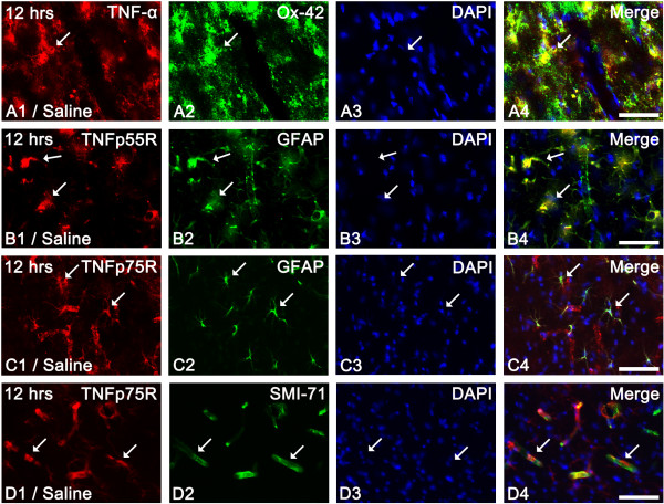
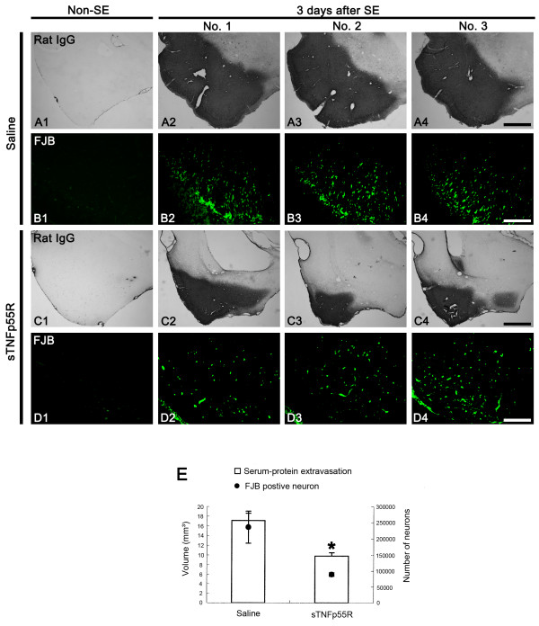
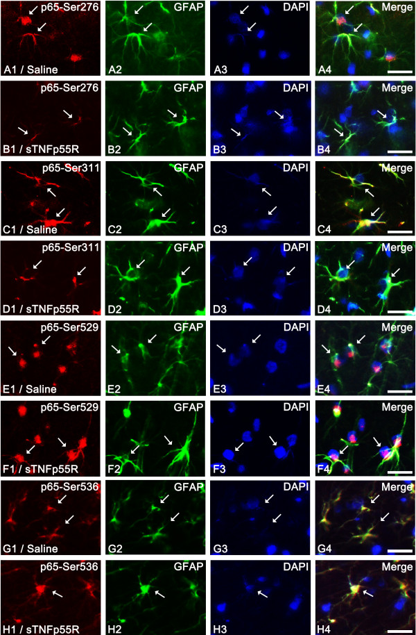
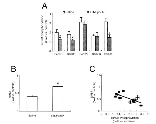
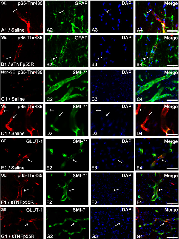
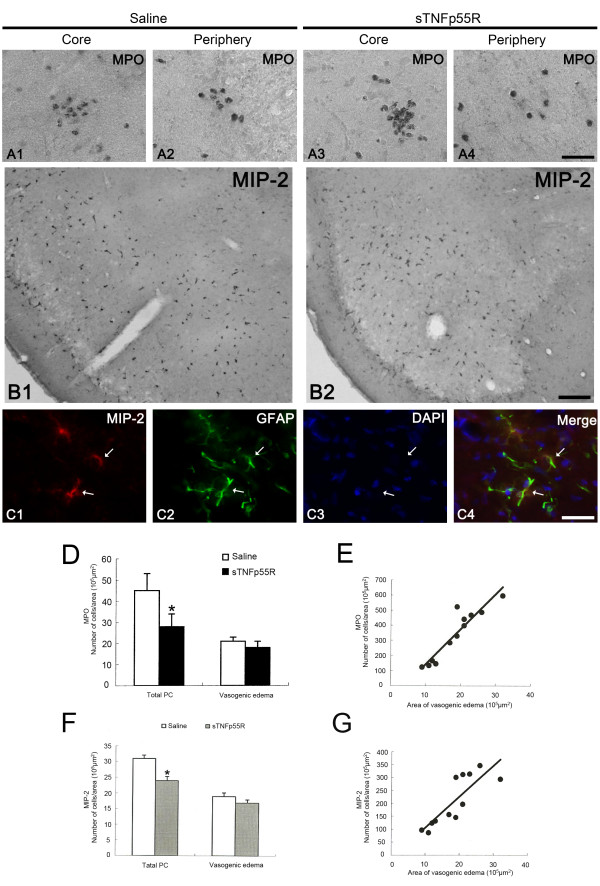
Results: Following SE, most activated microglia showed strong TNF-α immunoreactivity. In addition, TNF p75 receptor expression was detected in endothelial cells as well as astrocytes. In addition, only p65-Thr435 phosphorylation was increased in endothelial cells accompanied by SMI-71 expression (an endothelial barrier antigen). Neutralization of TNF-α by soluble TNF p55 receptor (sTNFp55R) infusion attenuated SE-induced vasogenic edema and neuronal damages via inhibition of p65-Thr435 phosphorylation in endothelial cells. Furthermore, sTNFp55R infusion reduced SE-induced neutrophil infiltration in the PC.

Conclusion: These findings suggest that impairments of endothelial cell functions via TNF-α-mediated p65-Thr 485 NF-κB phosphorylation may be involved in SE-induced vasogenic edema. Subsequently, vasogenic edema results in extensive neutrophil infiltration and neuronal-astroglial loss.


Figures

Figure 1
Figure 1
Expression of TNF-α and TNF receptor in the PC 12 hr-post SE. (A) TNF-α immunoreactivity in Ox-42-positive microglia (arrows). (B) TNFp55R expression in astrocytes (arrows). (C-D) TNFp75R expression in astrocytes as well as endothelial cells (arrows). Bar = 25 (A-D) μm.
Figure 2
Figure 2
Effect of sTNFp55R infusion on SE-induced serum-protein extravasation and neuronal damage. (A-D) Serum-protein extravasation and FJB-positive neuronal damages in the PC 3 days after SE. Compared to saline-infused animals, serum-protein extravasation and FJB-positive neuronal damage is markedly ameliorated in sTNFp55R-infused animals. Bars = 400 (A and C) and 50 (B and D) μm. (E) Quantitative analyses of serum-protein extravasation and FJB-positive neuronal damage in the PC 3 days after SE (mean ± S.E.M). Significant differences from saline-treated animals, *p < 0.05.
Figure 3
Figure 3
Effect of sTNFp55R infusion on NF-κB phosphorylation in astrocytes 12 hr after SE. In 12 hr-post SE animals of the saline-infused group (A, C, E and G), astrocytes show p65-Ser276 (A), p65-Ser311 (C), p65-Ser529 (E), and p65-Ser536 (G) phosphorylation (arrows). sTNFp55R infusion (B, D, F and H) effectively reduces p65-Ser276 (B) and p65-Ser311 (D) phosphorylation, while it does not affect p65-Ser529 (F) and p65-Ser536 (H) phosphorylation (arrows). Bar = 12.5 μm.
Figure 4
Figure 4
Quantitative analyses of the effect of sTNFp55R infusion on NF-κB phosphorylation and SMI-71 expression. (A) Quantitative analysis of NF-κB phosphorylation 12 hr after SE (mean ± S.E.M). Significant differences from saline-infused animals, *p < 0.05. (B) Quantitative analysis of SMI-71 expression 1 day after SE (mean ± S.E.M). Significant differences from saline-infused animals, *p < 0.05. (C) Linear regression analysis between p65-Thr435 phosphorylation and SMI-71 in the PC following SE.
Figure 5
Figure 5
Effect of sTNFp55R infusion on p65-Thr435 phosphorylation in endothelial cells following SE. (A-B) Inhibition of p65-Thr435 phosphorylation by sTNFp55R infusion 12 hr after SE. p65-Thr435 phosphorylation is rarely observed in astrocytes (arrows). (C) Endothelial p65-Thr435 phosphorylation in non-SE animals. (D-E) Endothelial p65-Thr435 phosphorylation in saline-infused animals 1 day after SE. p65-Thr435 phosphorylation is enhanced, while SMI-71 expression is reduced in GLUT-1-positive endothelial cells (arrows). (F-G) Endothelial p65-Thr435 phosphorylation in sTNFp55R-infused animal 1 day after SE. sTNFp55R infusion effectively reduces p65-Thr435 and preserves SMI-71 expression in GLUT-1-positive endothelial cells (arrows). Bars = 12.5 (A-D) and 25 (E-G) μm.
Figure 6
Figure 6
Effect of sTNFp55R infusion on neutrophil infiltration and MIP-2 expression following SE. (A) Neutrophil infiltration in vasogenic edema lesion 1 day after SE. (B) MIP-2 expression in the PC 1 day after SE. (C) Astroglial expression of MIP-2 (arrows). Bars = 12.5 (A and C) and 150 (B) μm. (D) Quantitative analysis of neutrophil infiltration 1 day after SE (mean ± S.E.M). Significant differences from saline-infused animals, *p < 0.05. (E) Linear regression analysis between the number of infiltrated neutrophils/area in the vasogenic edema region and the area of vasogenic edema in the PC. (F) Quantitative analysis of the number of MIP-2 positive cells per the unit area of vasogenic edema 1 day after SE (mean ± S.E.M). There is no difference in the number of MIP-2-positive cells per unit area of vasogenic edema between sTNFp55R-infused animals and saline-infused animals. (G) Linear regression analysis between the number of MIP-2 positive cells per unit area in vasogenic edema region and the area of vasogenic edema in the PC.

References

    1. DeLorenzo RJ, Pellock JM, Towne AR, Boggs JG. Epidemiology of status epilepticus. J Clin Neurophysiol. 1995;12:316–325. - PubMed
    1. Rice AC, DeLorenzo RJ. NMDA receptor activation during status epilepticus is required for the development of epilepsy. Brain Res. 1998;782:240–247. doi: 10.1016/S0006-8993(97)01285-7. - DOI - PubMed
    1. Stewart LS, Persinger MA. Ketamine Prevents Learning Impairment When Administered Immediately after Status Epilepticus Onset. Epilepsy Behav. 2001;2:585–591. doi: 10.1006/ebeh.2001.0272. - DOI - PubMed
    1. Gale K. Subcortical structures and pathways involved in convulsive seizure generation. J Clin Neurophysiol. 1992;9:264–277. doi: 10.1097/00004691-199204010-00007. - DOI - PubMed
    1. Sheen SH, Kim JE, Ryu HJ, Yang Y, Choi KC, Kang TC. Decrease in dystrophin expression prior to disruption of brain-blood barrier within the rat piriform cortex following status epilepticus. Brain Res. 2010;1369:173–183. - PubMed

Publication types

MeSH terms