Review
. 2012 Feb;33(1):71-108.
doi: 10.1210/er.2011-0013. Epub 2012 Jan 12.

Emerging role of mast cells and macrophages in cardiovascular and metabolic diseases

Affiliations
Review

Emerging role of mast cells and macrophages in cardiovascular and metabolic diseases

Jia-Ming Xu et al. Endocr Rev. 2012 Feb.

Abstract

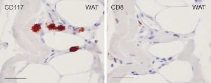
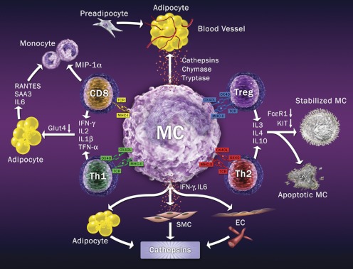
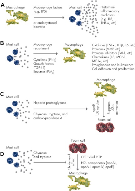
Mast cells are essential in allergic immune responses. Recent discoveries have revealed their direct participation in cardiovascular diseases and metabolic disorders. Although more sophisticated mechanisms are still unknown, data from animal studies suggest that mast cells act similarly to macrophages and other inflammatory cells and contribute to human diseases through cell-cell interactions and the release of proinflammatory cytokines, chemokines, and proteases to induce inflammatory cell recruitment, cell apoptosis, angiogenesis, and matrix protein remodeling. Reduced cardiovascular complications and improved metabolic symptoms in animals receiving over-the-counter antiallergy medications that stabilize mast cells open another era of mast cell biology and bring new hope to human patients suffering from these conditions.


Figures

Figure 1.
Figure 1.
MC activation pathways. Various extracellular soluble ligands can bind to corresponding receptors on MC surfaces, followed by MC activation or degranulation as indicated. An active MC is shown on the lower right.
Figure 2.
Figure 2.
MC and cathepsin S expression in human atherosclerotic lesions. Mouse antihuman tryptase monoclonal antibody (1:1500; Chemicon International, Temecula, CA) and rabbit antihuman cathepsin S polyclonal antibody (1:100) (60) detected tryptase-positive MC and cathepsin S expression on lumen endothelium and subendothelium compartment in human carotid artery fatty streak (100 μm), and both are colocalized in advanced atherosclerotic lesions (scale, 250 μm). Framed areas are shown enlarged in the right panels (scale, 100 μm). No cathepsin S or MC tryptase immunoreactivities were detected in normal aortas (scale, 100 μm). Frozen sections (6 μm) were used for immunostaining.
Figure 3.
Figure 3.
MC functions in atherosclerosis. MC produce cytokines, chemokines, chymase, tryptase, cathepsins, and other inflammatory mediators to promote angiogenesis, vascular-cell apoptosis, blood-borne inflammatory cell adhesion and recruitment, foam-cell formation, lipid degradation, Ang-II generation, and activation of zymogen, procytokine, and latent growth factors.
Figure 4.
Figure 4.
Localization of MC, macrophages, and CD8+ T cells in ruptured human AAA lesions. Mouse antihuman tryptase monoclonal antibody (1:1500; Chemicon International), mouse antihuman CD68 monoclonal antibody (1:60; Dako, Glostrup, Denmark), and mouse antihuman CD8 monoclonal antibody (1:75; Abcam, Cambridge, MA) detected tryptase+ MC, CD68+ macrophages, and CD8+ T cells in ruptured human AAA media (A, scale, 100 μm), adventitia (B, scale, 100 μm), and vasa vasorum surrounding the adventitia (C, scale, 50 μm). Paraffin sections (5 μm) were used.
Figure 5.
Figure 5.
MC and CD8+ T cells in mouse WAT. CD117+ MC (left, rat antimouse CD117 monoclonal antibody, 1:40; eBioscience, San Diego, CA) and CD8+ T cells (right, rabbit antimouse CD8 monoclonal antibody, 1:300; Abcam) appear in the same locations in WAT from ob/ob mice. Scale, 50 μm.
Figure 6.
Figure 6.
MC and T cell interaction model. MC use MHC class-I to interact with T-cell receptor on CD8+ T cells, to stimulate inflammatory cytokines and monocyte chemokine MIP-1α expression. MC also activate Th1 cells via MHC class II and OX40L to stimulate Th1 cell inflammatory cytokine expression, which activates adipocytes. MC may activate Treg and Th2 cells using MHC class II and OX40L. Both Treg and Th2 may release IL-3, IL-4, and IL-10 to stabilize MC by decreasing MC FcεR1 and KIT expression or directly causing MC apoptosis. MC also produce proteases to promote angiogenesis and adipogenesis in adipose tissues or release inflammatory cytokines to induce vascular cell and adipocyte cathepsin expression, angiogenesis, and adipogenesis.
Figure 7.
Figure 7.
MC–macrophage interaction. A, Macrophages release macrophage factors or endocytosed bacteria (e.g., C. pneumoniae) to activate MC. B, MC contribute to macrophage recruitment or release cytokines, growth factors, or enzymes to activate macrophages. C, MC release heparin proteoglycans or proteases to target apoB or nuclear receptor LXRα, thereby promoting macrophage LDL uptake and foam-cell formation. D, MC also release proteases to degrade the lipid transfer proteins CETP and PLTP, or to target directly HDL pre-β-migrating particles containing apoA-I, apoA-II, apoA-IV, and apoE, thereby inhibiting macrophage foam cell cholesterol efflux.

References

    1. Okayama Y, Kawakami T. 2006. Development, migration, and survival of mast cells. Immunol Res 34:97–115 - PMC - PubMed
    1. Galli SJ, Kalesnikoff J, Grimbaldeston MA, Piliponsky AM, Williams CM, Tsai M. 2005. Mast cells as “tunable” effector and immunoregulatory cells: recent advances. Annu Rev Immunol 23:749–786 - PubMed
    1. Hallgren J, Jones TG, Abonia JP, Xing W, Humbles A, Austen KF, Gurish MF. 2007. Pulmonary CXCR2 regulates VCAM-1 and antigen-induced recruitment of mast cell progenitors. Proc Natl Acad Sci USA 104:20478–20483 - PMC - PubMed
    1. Haley KJ, Lilly CM, Yang JH, Feng Y, Kennedy SP, Turi TG, Thompson JF, Sukhova GH, Libby P, Lee RT. 2000. Overexpression of eotaxin and the CCR3 receptor in human atherosclerosis: using genomic technology to identify a potential novel pathway of vascular inflammation. Circulation 102:2185–2189 - PubMed
    1. Irani AA, Schechter NM, Craig SS, DeBlois G, Schwartz LB. 1986. Two types of human mast cells that have distinct neutral protease compositions. Proc Natl Acad Sci USA 83:4464–4468 - PMC - PubMed

Publication types

MeSH terms