. 2012 Apr;142(4):938-46.
doi: 10.1053/j.gastro.2011.12.044. Epub 2012 Jan 10.

Autophagy releases lipid that promotes fibrogenesis by activated hepatic stellate cells in mice and in human tissues

Affiliations

Autophagy releases lipid that promotes fibrogenesis by activated hepatic stellate cells in mice and in human tissues

Virginia Hernández-Gea et al. Gastroenterology. 2012 Apr.

Abstract

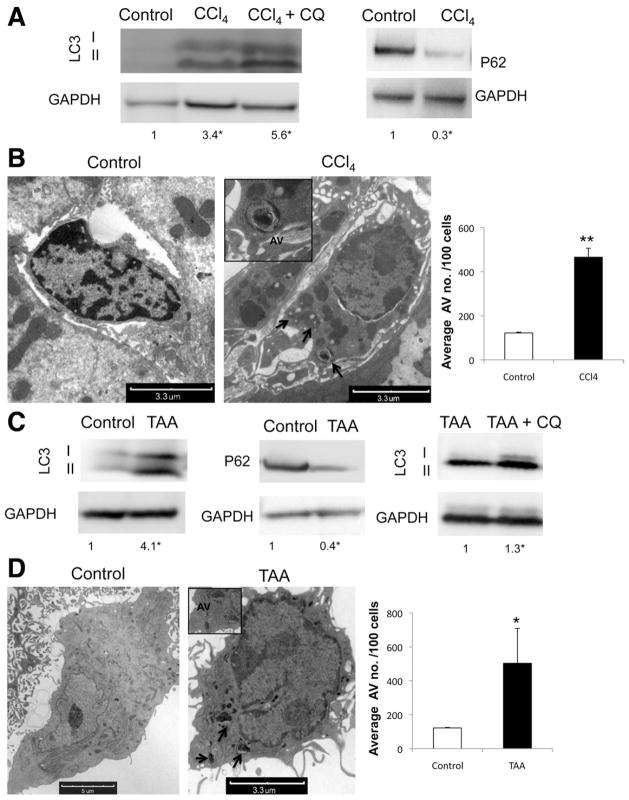
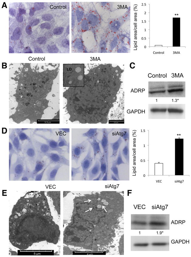
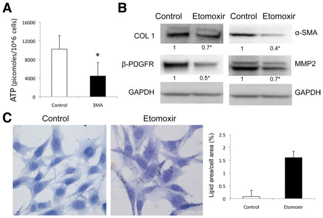
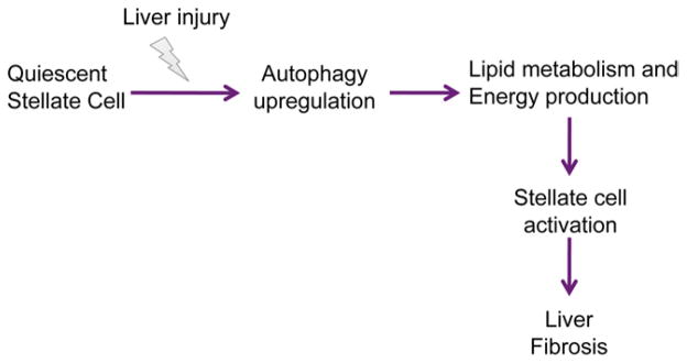
Background & aims: The pathogenesis of liver fibrosis involves activation of hepatic stellate cells, which is associated with depletion of intracellular lipid droplets. When hepatocytes undergo autophagy, intracellular lipids are degraded in lysosomes. We investigated whether autophagy also promotes loss of lipids in hepatic stellate cells to provide energy for their activation and extended these findings to other fibrogenic cells.

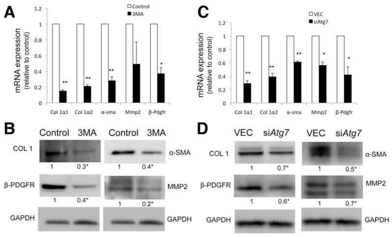
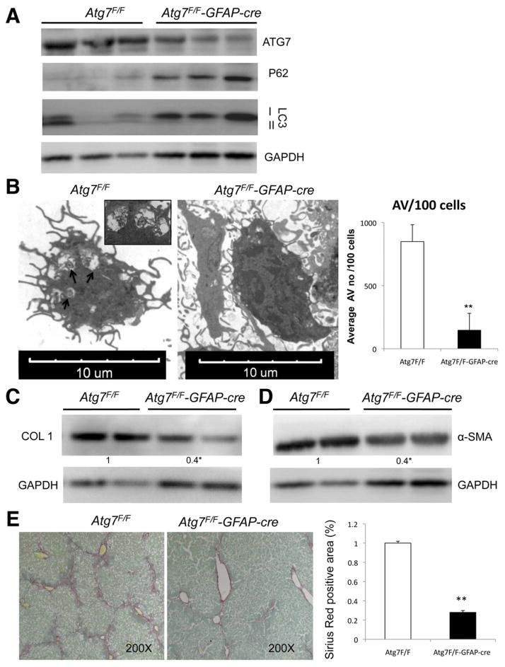
Methods: We analyzed hepatic stellate cells from C57BL/6 wild-type, Atg7(F/F), and Atg7(F/F)-GFAP-Cre mice, as well as the mouse stellate cell line JS1. Fibrosis was induced in mice using CCl(4) or thioacetamide (TAA); liver tissues and stellate cells were analyzed. Autophagy was blocked in fibrogenic cells from liver and other tissues using small interfering RNAs against Atg5 or Atg7 and chemical antagonists. Human pulmonary fibroblasts were isolated from samples of lung tissue from patients with idiopathic pulmonary fibrosis or from healthy donors.

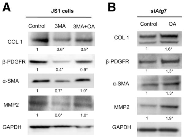
Results: In mice, induction of liver injury with CCl(4) or TAA increased levels of autophagy. We also observed features of autophagy in activated stellate cells within injured human liver tissue. Loss of autophagic function in cultured mouse stellate cells and in mice following injury reduced fibrogenesis and matrix accumulation; this effect was partially overcome by providing oleic acid as an energy substrate. Autophagy also regulated expression of fibrogenic genes in embryonic, lung, and renal fibroblasts.

Conclusions: Autophagy of activated stellate cells is required for hepatic fibrogenesis in mice. Selective reduction of autophagic activity in fibrogenic cells in liver and other tissues might be used to treat patients with fibrotic diseases.


Conflict of interest statement

Conflicts of interest

The authors disclose no conflicts.

Figures

Figure 1
Figure 1
Autophagy is up-regulated in stellate cells after liver injury in vivo. (A and C) Immunoblots of stellate cells isolated from wild-type mice after acute liver injury induction with (A) CCl4 and (C) TAA with and without addition of CQ showing an increase of LC3II conversion and decrease in P62. (B and D) Electron micrographs of whole liver tissue, showing stellate cells after (B) CCl4 and (D) TAA treatment. Arrows indicate AV. (Right) Electron micrograph quantification of AV number per 100 cells (P < .05, ★★P < .001; error bars indicate SEM). Protein ratios (normalized to GAPDH) were used to quantify fold change relative to control and are shown below each blot. Data represent the mean value of at least 3 experiments (P < .05), and 3 animals per condition were used in this experiment.
Figure 2
Figure 2
Inhibition of autophagy decreases fibrogenesis. (A and C) Col 1α1, Col 1α2, α-sma, Mmp2, and β-Pdgfr messenger RNA (quantitative reverse-transcription polymerase chain reaction analysis) and (B and D) protein expression (immunoblot) in JS1 cells (A and B) after 24-hour treatment with 3-MA or (C and D) 8 days after transduction with siAtg7 lentivirus. All figures are representative of at least 3 independent experiments. Messenger RNA is expressed as fold change relative to control (P < .05, ★★P < .001; error bars indicate SEM). Protein ratios (normalized to GAPDH) were used to quantify fold change relative to control and are shown below each blot. Data represent the mean value of at least 3 experiments (P < .05).
Figure 3
Figure 3
Autophagy regulates stellate cell activation and fibrosis in vivo. (A) Immunoblots of stellate cells protein isolated from Atg7F/F and Atg7F/F-GFAP-cre mice showing decreased expression of ATG7, LC3-II, and increased P62. (B) Electron micrographs of stellate cells isolated from Atg7F/F and Atg7F/F-GFAP-cre mice and AV quantification (right) depict a significant decrease of AV in transgenic animals. Arrows indicate AV. (C and D) Immunoblots for (C) collagen type I and α-SMA in isolated stellate cells from Atg7F/F and Atg7F/F-GFAP-cre mice after chronic liver injury with CCl4. (E) Whole liver sections after chronic liver injury with CCl4 were stained for Sirius Red. (Right) Quantification of Sirius Red–positive area. P < .05, ★★P < .001. Error bars indicate SEM. Protein ratios (normalized to GAPDH) were used to quantify fold change relative to control and are shown below each blot. Data represent the mean value of at least 3 experiments (P < .05) and a total of 18 animals: 9 Atg7F/F and 9 Atg7F/F-GFAP-cre.
Figure 4
Figure 4
Autophagy deficiency in stellate cells leads to LD accumulation. Lipid content analysis in JS1 cells (AC) treated with 3-MA or (DF) transduced with siAtg7. (A and D) ORO staining and (right) quantification of ORO-stained area (P < .05, ★★P < .001). Error bars indicate SEM. (B and E) Electron micrographs showing neutral LDs (arrows) and (C and F) immunoblots for ADRP (error bars indicate SEM). Protein ratios (normalized to GAPDH) were used to quantify fold change relative to control and are shown below each blot. Data represent the mean value of at least 3 experiments (P < .05).
Figure 5
Figure 5
β-oxidation regulates stellate cell activation. (A) Autophagy was inhibited in JS1 cells with 3-MA, and ATP content was determined at 12 hours after treatment. (B and C) β-oxidation was blocked with etomoxir in JS1 cells after 12-hour expression of COL1, α-SMA, and β-PDGFR was determined by (B) immunoblot analysis and (C) LD content was examined by ORO staining (right, quantification of ORO-stained area). Levels of ATP are expressed in picomoles per 106 cells. Error bars indicate SEM. Protein ratios (normalized to GAPDH or tubulin) were used to quantify fold change relative to control and are shown below each blot. Data represent the mean value of at least 3 experiments (P < .05).
Figure 6
Figure 6
Simplified model depicting the role of autophagy in stellate cell activation and fibrogenesis. The response of stellate cells to hepatic injury involves the up-regulation of autophagy, which fuels stellate cell activation, in part through its role in lipid breakdown and energy production, which drives the fibrogenic response of the liver.
Figure 7
Figure 7
Decrease in fibrogenesis caused by autophagy inhibition can be rescued by exogenous FFAs. JS1 cells were treated with (A) 3-MA or transduced with (B) siAtg7 and supplemented with OA conjugated to BSA or with BSA alone. Expression of COL 1, α-SMA, MMP2, and β-PDGFR was determined by (A and B) immunoblot analysis. Protein ratios (normalized to GAPDH) were used to quantify fold change relative to control and are shown below each blot. Data represent the mean value of at least 3 experiments (P < .05).

References

    1. Mehal WZ, Iredale J, Friedman SL. Scraping fibrosis: expressway to the core of fibrosis. Nat Med. 2011;17:552–553. - PMC - PubMed
    1. Wynn TA. Cellular and molecular mechanisms of fibrosis. J Pathol. 2008;214:199–210. - PMC - PubMed
    1. Friedman SL. Mechanisms of hepatic fibrogenesis. Gastroenterology. 2008;134:1655–1669. - PMC - PubMed
    1. Friedman SL. Hepatic stellate cells: protean, multifunctional, and enigmatic cells of the liver. Physiol Rev. 2008;88:125–172. - PMC - PubMed
    1. Blaner WS, O’Byrne SM, Wongsiriroj N, et al. Hepatic stellate cell lipid droplets: a specialized lipid droplet for retinoid storage. Biochim Biophys Acta. 2009;1791:467–473. - PMC - PubMed

Publication types

MeSH terms