

High-contrast en bloc staining of neuronal tissue for field emission scanning electron microscopy
- PMID: 22240582
- PMCID: PMC3701260
- DOI: 10.1038/nprot.2011.439
High-contrast en bloc staining of neuronal tissue for field emission scanning electron microscopy
Abstract
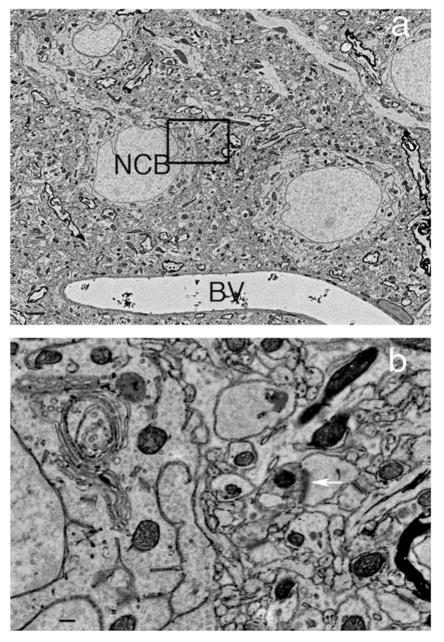
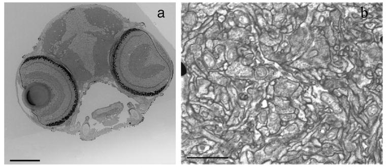
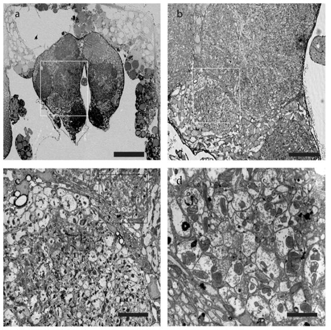
Conventional heavy metal poststaining methods on thin sections lend contrast but often cause contamination. To avoid this problem, we tested several en bloc staining techniques to contrast tissue in serial sections mounted on solid substrates for examination by field emission scanning electron microscopy (FESEM). Because FESEM section imaging requires that specimens have higher contrast and greater electrical conductivity than transmission electron microscopy (TEM) samples, our technique uses osmium impregnation (OTO) to make the samples conductive while heavily staining membranes for segmentation studies. Combining this step with other classic heavy metal en bloc stains, including uranyl acetate (UA), lead aspartate, copper sulfate and lead citrate, produced clean, highly contrasted TEM and scanning electron microscopy (SEM) samples of insect, fish and mammalian nervous systems. This protocol takes 7-15 d to prepare resin-embedded tissue, cut sections and produce serial section images.
Conflict of interest statement
The authors of this manuscript declare that they have no competing financial interests.
Figures
References
-
- Gay H, Anderson TF. Serial sections for electron microscopy. Science. 1954;120:1071–3. - PubMed
-
- Stevens JK, Davis TL, Friedman N, Sterling P. A systematic approach to reconstructing microcircuitry by electron microscopy of serial sections. Brain Res. 1980;3:265–93. - PubMed
-
- Blow N. Following the wires. Nat Methods. 2007;4:975–981.
Publication types
MeSH terms
Substances
Grants and funding
LinkOut - more resources
Full Text Sources
Other Literature Sources
Molecular Biology Databases
Research Materials
