. 2012 Jan 12;7(2):235-46.
doi: 10.1038/nprot.2011.437.

Isolation and characterization of mouse and human esophageal epithelial cells in 3D organotypic culture

Affiliations

Isolation and characterization of mouse and human esophageal epithelial cells in 3D organotypic culture

Jiri Kalabis et al. Nat Protoc. .

Abstract

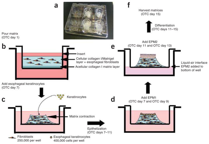
This protocol describes the isolation and characterization of mouse and human esophageal epithelial cells and the application of 3D organotypic culture (OTC), a form of tissue engineering. This model system permits the interrogation of mechanisms underlying epithelial-stromal interactions. We provide guidelines for isolating and cultivating several sources of epithelial cells and fibroblasts, as well as genetic manipulation of these cell types, as a prelude to their integration into OTC. The protocol includes a number of important applications, including histology, immunohistochemistry/immunofluorescence, genetic modification of epithelial cells and fibroblasts with retroviral and lentiviral vectors for overexpression of genes or RNA interference strategies, confocal imaging, laser capture microdissection, RNA microarrays of individual cellular compartments and protein-based assays. The OTC (3D) culture protocol takes 15 d to perform.


Conflict of interest statement

COMPETING FINANCIAL INTERESTS The authors declare no competing financial interests.

Figures

Figure 1
Figure 1
Establishment of OTC on inserts placed on plates (organogenesis). (a) Inserts are placed on plates. (b) Initially, there is placement of an acellular collagen matrix on the bottom of an insert, followed by the addition of a layer of esophageal fibroblasts embedded in bovine collagen type I. These two layers are cultured initially for 7 d, thereby allowing for fibroblast-mediated constriction of the collagen matrix. (c) On day 7, the epithelial cells are seeded on the surface of the contracted matrix. (d,e) The medium of the OTC is changed every 2 d (d) and the epithelium is exposed to air to create a liquid-air interface (e), thereby promoting epithelial stratification and differentiation. (f) Finally, on day 15, the resulting OTC is harvested for histology and other applications such as RNA/protein analyses.
Figure 2
Figure 2
The use of OTC for LCM. Specific cell populations (e.g., highlighted invading epithelial cells or regions of fibroblasts in the matrix) may be isolated using LCM from frozen sections. Scale bar, 50 μm.
Figure 3
Figure 3
Detection of side population of esophageal epithelial cells. (a) Forward (FSC-A) and side (SSC-A) scatter of primary esophageal epithelial cells. Single cells are gated (usually 200–400 FCS-A and 100–400 SSC-A). (b) Live cells gated as PI intensity below 101.3 (PI was excited at 488 nm and its emission was measured in a logarithmic scale through a band-pass filter of 630/22A). (c) Verapamil-treated cells show diminished side population fraction. (d) Side population cells (tail of cells with Hoechst red below 300 and Hoechst blue below 400) and side population–negative cells (Hoechst red above 300, Hoechst blue over 400). Modified from ref. with permission.
Figure 4
Figure 4
Morphology of esophageal OTC derived from mouse cells and immunohistological detection of proliferation and differentiation markers. (a) SP cells (103) from GFP+ mice mixed with 3 × 10⁴ unsorted GFP cells form a stratified epithelium. (b) A ×400 magnification of the same figure as in a. (cf) Immunohistochemical staining for Ki-67 (c), CK14 (d), CK4 (e) and CK13 (f). Original magnification, ×400. (g) Two-photon confocal microscopy on day 12 after seeding 103 NSP or transit-amplifying cells isolated from GFP+ mice (green) with DAPI+ nuclei (blue); top view, z-stack, 53.8 μm in 0.78-μm increments. (h) Two-photon confocal microscopy of the complete stratified epithelium on day 12 after seeding 103 SP cells isolated from GFP+ mice (green) with DAPI+ nuclei (blue); top view, z-stack, 60 μm in 1-μm increments. Arrowheads indicate basement membrane. Modified from ref. with permission. Scale bars, 25 μm.
Figure 5
Figure 5
Morphology of OTC derived from immortalized human keratinocytes (EPC2-hTERT). (a,b) Complete stratified epithelium with proliferative basal and differentiated suprabasal cells. Notch inhibition impairs squamous differentiation of esophageal epithelia reconstituted in 3D OTC. EPC2-hTERT cells were grown in the presence or absence of 1 μmol per liter compound E, a γ-secretase inhibitor (GSI) or retroviral DNMAML1 and subjected to histology. (b) Immunofluorescence for CK14 (red) and involucrin (green). Ep., epithelium; Str., stroma. Modified from ref. with permission. Scale bars, 50 μm.

References

    1. Al Yassin TM, Toner PG. Fine structure of squamous epithelium and submucosal glands of human oesophagus. J Anat. 1977;123:705–721. - PMC - PubMed
    1. Moll R, Franke WW, Schiller DL, Geiger B, Krepler R. The catalog of human cytokeratins: patterns of expression in normal epithelia, tumors and cultured cells. Cell. 1982;31:11–24. - PubMed
    1. Fuchs E. Epidermal differentiation: the bare essentials. J Cell Biol. 1990;111:2807–2814. - PMC - PubMed
    1. Grace MP, Kim KH, True LD, Fuchs E. Keratin expression in normal esophageal epithelium and squamous cell carcinoma of the esophagus. Cancer Res. 1985;45:841–846. - PubMed
    1. Lloyd C, et al. The basal keratin network of stratified squamous epithelia: defining K15 function in the absence of K14. J Cell Biol. 1995;129:1329–1344. - PMC - PubMed

Publication types