. 2012 Jan 31;106(3):496-507.
doi: 10.1038/bjc.2011.577. Epub 2012 Jan 12.

Combination of a fusogenic glycoprotein, pro-drug activation and oncolytic HSV as an intravesical therapy for superficial bladder cancer

Affiliations

Combination of a fusogenic glycoprotein, pro-drug activation and oncolytic HSV as an intravesical therapy for superficial bladder cancer

G R Simpson et al. Br J Cancer. .

Abstract

Background: There are still no effective treatments for superficial bladder cancer (SBC)/non-muscle invasive bladder cancer. Following treatment, 20% of patients still develop metastatic disease. Superficial bladder cancer is often multifocal, has high recurrences after surgical resection and recurs after intravesical live Bacillus Calmette-Guérin. Oncovex(GALV/CD), an oncolytic herpes simplex virus-1, has shown enhanced local tumour control by combining oncolysis with the expression of a highly potent pro-drug activating gene and the fusogenic glycoprotein.

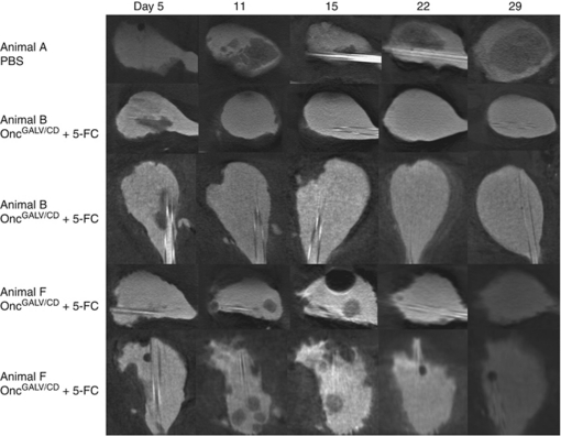
Methods: In vitro fusion/prodrug/apoptotic cell-based assays. In vivo orthotopic bladder tumour model, visualised by computed microtomography.

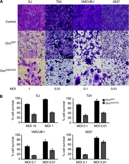
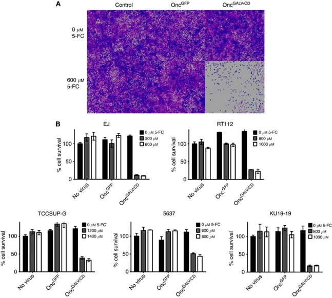
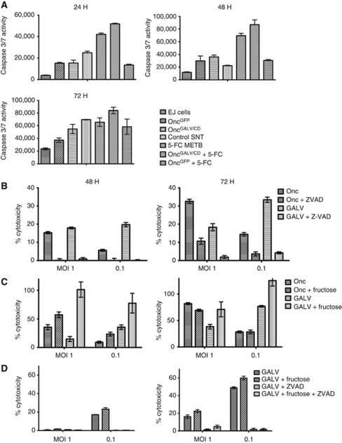
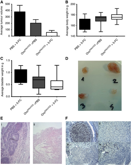
Results: Treatment of seven human bladder carcinoma cell lines with the virus resulted in tumour cell killing through oncolysis, pro-drug activation and glycoprotein fusion. Oncovex(GALV/CD) and mitomycin C showed a synergistic effect, whereas the co-administration with cisplatin or gemcitabine showed an antagonistic effect in vitro. Transitional cell cancer (TCC) cells follow an apoptotic cell death pathway after infection with Oncovex(GALV/CD) with or without 5-FC. In vivo results showed that intravesical treatment with Oncovex(GALV/CD) + prodrug (5-FC) reduced the average tumour volume by over 95% compared with controls.

Discussion: Our in vitro and in vivo results indicate that Oncovex(GALV/CD) can improve local tumour control within the bladder, and potentially alter its natural history.


Conflict of interest statement

We would also like to declare that Toby Price, and Robert S Coffin work for a commercial company Biovex Inc.

Figures

Figure 1
Figure 1
Fusogenic glycoprotein cytotoxic killing of bladder TCC cells. EJ, T24 VMCUB-I and 5637 cells were infected with OncovexGFP or OncoVexGALV/CD at various MOIs and incubated at 37 °C/5% CO2 for 48 h, then assayed by crystal violet staining (A) or MTS assay (B) (Promega). Average cell survival was calculated as a percentage compared with untreated cells (Unpaired student t-test comparing GFP Vs GALV/CD P-values: EJ (MOI 10 P<0.000, MOI 1 P<0.000), T24 (MOI 0.1 P<0.000, 0.01 P<0.000), VMCUMB-I (MOI 0.1 P<0.000, 0.01 P<0.000), 5637 (MOI 0.1 P<0.000, 0.01 P<0.000).
Figure 2
Figure 2
Prodrug activation in TCC cells infected OncovexGALV/CD. EJ, RT112, TCCSUP-G, 5637, and KU19-19 cells were infected with OncovexGFP and OncovexGALV/CD at MOI of 0.1 and no virus control. After 30 min at 37 °C/5% CO2, the virus was removed, and 1 ml of FGM containing 5-FC (C4H4FN2O; Sigma) at different concentrations (0-1400  μmol l–1) was added and incubated for 48 h at 37 °C/5% CO2. The cell supernatants were then heat inactivated and added to 1 x 10⁴ fresh target cells and incubated at 37 °C/5% CO2 for 72 h and assayed by fixing and staining with glutaraldehyde/crystal violet (A) or MTS assay (Promega) (B). Average cell survival was calculated as a percentage compared with untreated cells (unpaired student t-test comparing GALV/CD+or - 5-FC P-values: EJ (300 μmol P<0.000, 600 μmol P<0.000), RT112 (800 μmol P<0.000, 1000 μmol P<0.000), TCCSUP-G 1200 μmol P<0.000, 1400 μmol P<0.000), 5637 800 μmol P<0.000, 1000 μmol P<0.000), KU19-19 (800 μmol P<0.000, 1000 μmol P<0.000).
Figure 3
Figure 3
Oncolytic HSV viral replication in the presence of viral glycoprotein, 5-FC and metabolites of 5-FC. (A) EJ cells were infected at various MOI (0.001 0.0001) with either OncovexGFP or OncovexGALV/CD and incubated at 37 °C for 48 h. Unpaired student t-test comparing GFP vs GALV/CD P-value (MOI 0.001) 0.000 (MOI 0.0001) 0.0002. (B) EJ cells were infected at various MOIs (0.1, 0.01, 0.001) with OncovexGALV/CD in the presence or absence of 5-FC and incubated at 37 °C for 48 h. + or – 5-FC P-value (MOI 0.1) 0.03 (MOI 0.01) 0.414 (MOI 0.001) 0.075. (C) EJ cells were infected at various MOIs (0.1, 0.01. 0.001) with OncovexGALV/CD in the presence or absence of metabolites of 5-FC and incubated at 37 °C for 48 h. The resulting virus stocks were titred on BHK cells using a standard plaque assay + or – metabolites of 5-FC P-value (MOI 0.1) 0.005 (MOI 0.01) 0.004 (MOI 0.001) 0.0001.
Figure 4
Figure 4
Bladder TCC cells follow apoptotic pathway after infection with OncovexGALV/CD. (A) EJ cells were infected with OncovexGFP or OncovexGALV/CD (MOI 1) with or without 5-FC (600 μM) or its metabolites for 24, 48, and 72 h. Caspase activity was detected using Caspase Glo 3/7 reagent (Promega). Unpaired student t-test comparing GFP vs GALV/CD P-value 24 h 0.8919 48 h 0.2596 72 h 0.061. (B) EJ cells were infected OncovexGALV/CD or OncovexGFP at MOIs of 1, 0.1 and then incubated with and without 50 uM Z-VAD-FMK (Sigma) for 48/72 h. After which, the supernatant of the samples were assayed for lactate dehydrogenase activity (LDH) a marker of cytotoxic cell death (Roche). (C) EJ cells were infected with OncovexGALV/CD or OncovexGFP at MOIs of 1, 0.1 and then incubated with and without 20 mM fructose and incubated for 48/72 h at 37 °C. LDH assays were carried out on sample supernatant. (D) EJ cells were infected with OncovexGALV/CD at MOIs of 1, 0.1 and then incubated with either 50 μM Z-VAD-FMK or 20 mM fructose or both and incubated/assayed as above (unpaired student t-test comparing P-value Onc vs Onc ZVAD, 48 h MOI 1 P>0.0000, MOI 0.1 P 0.0127, 72 h MOI 1 P>0.0000, MOI 0.1 P 0.000. GALV vs GALV ZVAD 48 h MOI 1 P>0.0000, MOI 0.1 P>0.0000, 72 h MOI 1 P>0.0001, MOI 0.1 P>0.0000. Onc vs Onc Fru, 48 h MOI 1 P0.0078, MOI 0.1 P 0.0032, 72 h MOI 1 0.0051, MOI 0.1 0.3229, GALV vs GALV Fru 48 h MOI 1 P>0.0015, MOI 0.1 P>0.0001, 72 h MOI 1 0.0595, MOI 0.1 0.0148).
Figure 5
Figure 5
Treatment of a rat orthotopic bladder tumour model with OncovexGALV/CD +5-FC. AY-27 HVEM cells were implanted into the bladders of female Fischer F344 rats at day 0. The tumour-bearing animals were assigned into three treatment groups either OncovexGALV/CD+5-FC (n=10), OncovexGALV/CD +PBS (n=10) or PBS+5-FC (control group) (n=8). Intravesical treatment of implanted tumours was carried out with virus (OncovexGALV/CD, 9e7 pfu) or control PBS on days 7, 14, and 21. Prodrug 5-FC (12.5 mg ml–1) or PBS was installed in the same manner on days 8, 9, 15, 16, 22, and 24 and the animals were killed on day 28. Their bladders were removed and assessed for tumour abundance. The harvested bladders were weighed, then opened up and the bladder surface, which macroscopically contained tumour was measured with a caliper. Average tumour volume was calculated by measuring length x width/2. (A) Average tumour volume (B) Average body weight (C) Average bladder weight (unpaired student t-test comparing tumour volume PBS vs GALV/CD +5-FC P=0.001, PBS GALV/CD- 5-FC vs GALV/CD +5-FC P=0.034, PBS vs GALV/CD −5-FC P=0.13. (D) Bladder seeded with tumour cells (no treatment) and removed at necropsy after 18 days (1, 2 and 3). Bladder 4 was a control in which bladder mucosa was conditioned (acid/alkali rinse) but no cells were seeded. (E) H&E stain of sections from bladder from animals A (i) and B (ii). (F) Ki67 stain (brown) of sections from bladder from animals A (i) and B (iii). No primary antibody control sections from animals A (ii) and B (iv).
Figure 6
Figure 6
Computed tomography imaging of an orthotopic bladder tumour model during treatment with OncovexGALV/CD + 5-FC. Intravesical treatment of implanted tumours was carried out with virus (OncovexGALV/CD, 9e7 pfu) (or Control PBS) on days 6, 12, and 18. Prodrug 5-FC (12.5 mg ml–1) was installed in the same manner on days 7, 8, 13, 14, 19, and 20 and the animals were killed on day 29. During the experiment, computed tomography imaging was carried out on days 5, 11, 15, 22, and 29.

References

    1. Adam L, Black PC, Kassouf W, Eve B, McConkey D, Munsell MF, Benedict WF, Dinney CP (2007) Adenoviral mediated interferon-alpha 2b gene therapy suppresses the pro-angiogenic effect of vascular endothelial growth factor in superficial bladder cancer. J Urol 177: 1900–1906 - PubMed
    1. Adusumilli PS, Chan MK, Chun YS, Hezel M, Chou TC, Rusch VW, Fong Y (2006) Cisplatin-induced GADD34 upregulation potentiates oncolytic viral therapy in the treatment of malignant pleural mesothelioma. Cancer Biol Ther 5: 48–53 - PMC - PubMed
    1. Andreansky SS, He B, Gillespie GY, Soroceanu L, Markert J, Chou J, Roizman B, Whitley RJ (1996) The application of genetically engineered herpes simplex viruses to the treatment of experimental brain tumors. Proc Natl Acad Sci USA 93: 11313–11318 - PMC - PubMed
    1. Anundi I, de GH (1989) Hypoxic liver cell death: critical Po2 and dependence of viability on glycolysis. Am J Physiol 257: G58–G64 - PubMed
    1. Bateman A, Bullough F, Murphy S, Emiliusen L, Lavillette D, Cosset FL, Cattaneo R, Russell SJ, Vile RG (2000) Fusogenic membrane glycoproteins as a novel class of genes for the local and immune-mediated control of tumor growth. Cancer Res 60: 1492–1497 - PubMed

MeSH terms