. 2012 Feb;26(2):281-91.
doi: 10.1210/me.2011-1299. Epub 2012 Jan 12.

Deletion of CaMKK2 from the liver lowers blood glucose and improves whole-body glucose tolerance in the mouse

Affiliations

Deletion of CaMKK2 from the liver lowers blood glucose and improves whole-body glucose tolerance in the mouse

Kristin A Anderson et al. Mol Endocrinol. 2012 Feb.

Abstract

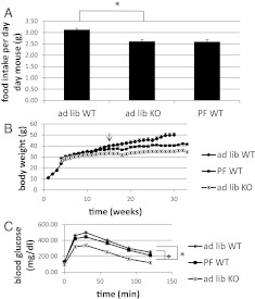
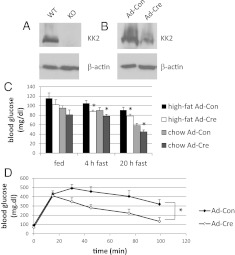
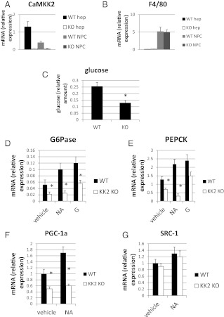
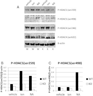
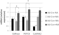
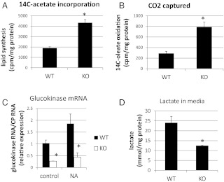
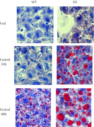
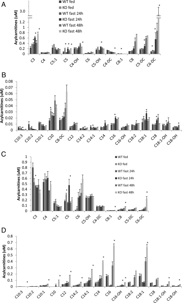
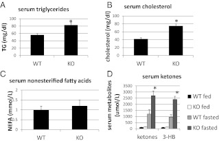
Ca(2+)/calmodulin-dependent protein kinase kinase 2 (CaMKK2) is a member of the Ca(2+)/CaM-dependent protein kinase family that is expressed abundantly in brain. Previous work has revealed that CaMKK2 knockout (CaMKK2 KO) mice eat less due to a central nervous system -signaling defect and are protected from diet-induced obesity, glucose intolerance, and insulin resistance. However, here we show that pair feeding of wild-type mice to match food consumption of CAMKK2 mice slows weight gain but fails to protect from diet-induced glucose intolerance, suggesting that other alterations in CaMKK2 KO mice are responsible for their improved glucose metabolism. CaMKK2 is shown to be expressed in liver and acute, specific reduction of the kinase in the liver of high-fat diet-fed CaMKK2(floxed) mice results in lowered blood glucose and improved glucose tolerance. Primary hepatocytes isolated from CaMKK2 KO mice produce less glucose and have decreased mRNA encoding peroxisome proliferator-activated receptor γ coactivator 1-α and the gluconeogenic enzymes glucose-6-phosphatase and phosphoenolpyruvate carboxykinase, and these mRNA fail to respond specifically to the stimulatory effect of catecholamine in a cell-autonomous manner. The mechanism responsible for suppressed gene induction in CaMKK2 KO hepatocytes may involve diminished phosphorylation of histone deacetylase 5, an event necessary in some contexts for derepression of the peroxisome proliferator-activated receptor γ coactivator 1-α promoter. Hepatocytes from CaMKK2 KO mice also show increased rates of de novo lipogenesis and fat oxidation. The changes in fat metabolism observed correlate with steatotic liver and altered acyl carnitine metabolomic profiles in CaMKK2 KO mice. Collectively, these results are consistent with suppressed catecholamine-induced induction of gluconeogenic gene expression in CaMKK2 KO mice that leads to improved whole-body glucose homeostasis despite the presence of increased hepatic fat content.


Figures

Fig. 1.
Fig. 1.
Pair feeding WT mice does not improve negative effects of high-fat diet on glucose homeostasis. A, Pair-fed WT (PF WT) mice were restricted daily to the amount of high-fat food consumed ad libitum by CaMKK2 KO (ad lib KO) mice. A second group of WT mice were given free access to food (ad lib WT). B, Body weight accumulation of ad lib and PF mice over the 30-wk time course of the experiment. Arrow indicates wk 14, at which point ad lib WT and PF WT body weight becomes statistically different. At wk 30, PF WT mice do not show significantly improved glucose tolerance (C) when compared with ad lib KO. (n = 14).
Fig. 2.
Fig. 2.
Liver-specific deletion of CaMKK2 lowers blood glucose and improves whole-body glucose tolerance. CaMKK2 protein is detected by Western blot in liver extract from WT mice (A). Exposure of chow or high-fat diet-fed CaMKK2floxed mice to adenovirus-cre (Ad-Cre) reduces CaMKK2 expression by approximately 50% relative to mice exposed to control virus (Ad-Con). Shown is one representative blot (B). Blood glucose of fed and fasted animals was determined 2 wk after viral transfer (C), and glucose tolerance (D) of high-fat diet-fed mice assayed (n = 5).
Fig. 3.
Fig. 3.
Basal and catecholamine-induced gluconeogenesis is suppressed in primary hepatocytes from CaMKK2 KO mice. Hepatocyte-enriched (hep) and NPC fractions were isolated from WT and CaMKK2 KO mouse liver that are enriched in CaMKK2 (A) and F4/80 (B) mRNA, respectively. C, After a 6-h incubation of hepatocytes in glucose-free media, glucose present in the growth media was quantified. D–G, After a 6-h incubation in glucose-free media the indicated mRNAs, G6Pase, PEPCK, PGC-1a, and SRC-1 were quantified. Included during the incubation was glucagon (G) or NA (n = 3).
Fig. 4.
Fig. 4.
A, Basal and catecholamine-induced phosphorylation of HDAC5 is suppressed in primary hepatocytes from CaMKK2 KO mice. Primary hepatocytes from WT and CaMKK2 KO mice incubated in hepatocyte media were treated with PBS (vehicle), 1 μm ionomycin (ion) for 5 min, or 10 μm NA for 10 min, after which protein extracts were made for PAGE and Western blotting with antibodies against P-HDAC5(ser259)/P-HDAC4(ser246), P-HDAC5(ser498)/P-HDAC4(ser632), and β-actin. W, Wild type; K, CaMKK2 KO hepatocyte samples, respectively. B and C, Signal was quantified by densitometry. Shown is one representative experiment (n = 3).
Fig. 5.
Fig. 5.
The catecholamine signaling defect in hepatocytes from CaMKK2 KO mice is cell autonomous. CaMKK2 was deleted from primary hepatocytes from CaMKK2floxed mice by adenovirus-mediated gene transfer of cre-recombinase (Ad-Cre). Some cells received instead control virus (Ad-Con). The cells were switched 24 h later to glucose-free media in the presence and absence of NA for 6 h, after which mRNA for G6Pase, PEPCK, and CaMKK2 was quantified (n = 3).
Fig. 6.
Fig. 6.
Fat metabolism in primary hepatocytes from CaMKK2 KO mice is enhanced. Hepatocytes from CaMKK2 KO mice show increased rates of de novo lipogenesis (A) and fatty acid oxidation (B). To determine glucokinase mRNA levels and lactate production, hepatocytes were cultured overnight in hepatocyte media with 25 mm glucose. The next morning control cells or those treated with NA for 5 h were harvested, and the quantity of glucokinase mRNA, normalized for cyclophilin, was determined by RT-PCR (C). Cell culture media were collected overnight, and lactate was quantified using L-Lactate Assay Kit (Eton Bioscience, Inc.) (panel D). [n = 3 (panel A), n = 5 (panel B), n = 3 (panel C), and n = 3 (panel D)].
Fig. 7.
Fig. 7.
Liver from CaMKK2 KO mice is steatotic. Liver tissue sections from WT and CaMKK2 KO mice from chow-fed mice in fed and fasted states were stained with hematoxylin and the neutral lipid dye, Oil Red O. Shown are representative images in which n = 10.
Fig. 8.
Fig. 8.
Liver and serum acyl carnitine profiles. Short (A and C) and medium and long-chain length acyl carnitines were quantified in liver (A and B) and serum (C and D) from WT and CaMKK2 KO mice (n = 5).
Fig. 9.
Fig. 9.
A–D, Serum triglyceride, cholesterol, and ketones are increased in serum from CaMKK2 KO chow-fed mice (n = 9). NEFA, Nonesterified fatty acid; TG, Triglyceride; 3-HB, 3-hydroxybutyrate.

References

    1. Muoio DM, Newgard CB. 2008. Mechanisms of disease: molecular and metabolic mechanisms of insulin resistance and β-cell failure in type 2 diabetes. Nat Rev Mol Cell Biol 9:193–205 - PubMed
    1. Beck-Nielsen H, Hother-Nielsen O, Staehr P. 2002. Is hepatic glucose production increased in type 2 diabetes mellitus? Curr Diab Rep 2:231–236 - PubMed
    1. Gaede P, Vedel P, Parving HH, Pedersen O. 1999. Intensified multifactorial intervention in patients with type 2 diabetes mellitus and microalbuminuria: the Steno type 2 randomised study. Lancet 353:617–622 - PubMed
    1. He L, Sabet A, Djedjos S, Miller R, Sun X, Hussain MA, Radovick S, Wondisford FE. 2009. Metformin and insulin suppress hepatic gluconeogenesis through phosphorylation of CREB binding protein. Cell 137:635–646 - PMC - PubMed
    1. Jeng CY, Sheu WH, Fuh MM, Chen YD, Reaven GM. 1994. Relationship between hepatic glucose production and fasting plasma glucose concentration in patients with NIDDM. Diabetes 43:1440–1444 - PubMed

Publication types

MeSH terms