

A comparison of intrascleral bleb height by anterior segment OCT using three different implants in deep sclerectomy
- PMID: 22241011
- PMCID: PMC3325577
- DOI: 10.1038/eye.2011.358
A comparison of intrascleral bleb height by anterior segment OCT using three different implants in deep sclerectomy
Abstract
Purpose: To compare intrascleral blebs characteristics after deep sclerectomy (DS) with three intrascleral implants using the Visante anterior segment optical coherence tomography.
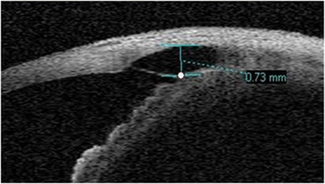
Methods: This is a cross-sectional study including 60 eyes of 51 patients that underwent DS with Sk-Gel, Esnoper, and Aquaflow implant. Intraocular pressure (IOP) measurement, slit-lamp examination, and Visante scans were performed the same day in all the patients. Visante scans were done through the intrascleral bleb at 45°, 90°, and 135° and the bleb height was measured.
Results: Sk-Gel was used in 19 eyes (31.66%), Esnoper in 22 eyes (36.66%), and Aquaflow in 19 eyes (31.66%). The median time lapsed from the surgery was 15.50 months 25th and 75th percentiles (p(25) 8.25; p(75) 20). The median IOP was 13 mm Hg (p(25) 10; p(75) 15), with no significant differences among implants (P = 0.232). Overall, the correlation between the scleral bleb height and the IOP was statistically significant at 45° (r=-0.359; P = 0.004), 90° (r = -0.410; P = 0.001), and 135° (r = -0.417; P = 0.001). However, Sk-Gel did not show any statistically significant correlation between the scleral height and IOP whereas the other two groups (Esnoper and Aquaflow) showed a significant correlation. There were no differences in the bleb height among implants.
Conclusion: There was a moderate inverse correlation between the scleral bleb height and the IOP measurement after DS with Esnoper and Aquaflow implants. There were no differences in bleb height among the three implants.
Figures
Similar articles
-
Two-year Results After Deep Sclerectomy With Nonabsorbable Uveoscleral Implant (Esnoper-Clip): Surgical Area Analysis Using Anterior Segment Optical Coherence Tomography.J Glaucoma. 2017 Oct;26(10):929-935. doi: 10.1097/IJG.0000000000000756. J Glaucoma. 2017. PMID: 28991150
-
Visante anterior segment optical coherence tomography analysis of morphologic changes after deep sclerectomy with intraoperative mitomycin-C and no implant use.J Glaucoma. 2014 Jan;23(1):e86-90. doi: 10.1097/IJG.0b013e31829ea2c8. J Glaucoma. 2014. PMID: 24370813
-
Comparison of bleb morphology between trabeculectomy and deep sclerectomy using a clinical grading scale and anterior segment optical coherence tomography.Clin Exp Ophthalmol. 2017 Sep;45(7):701-707. doi: 10.1111/ceo.12953. Epub 2017 May 9. Clin Exp Ophthalmol. 2017. PMID: 28371125
-
Anterior Segment Optical Coherence Tomography Imaging of Filtering Blebs after Deep Sclerectomy with Esnoper-Clip Implant: One-year Follow-up.J Curr Glaucoma Pract. 2014 Sep-Dec;8(3):91-5. doi: 10.5005/jp-journals-10008-1169. Epub 2015 Jan 15. J Curr Glaucoma Pract. 2014. PMID: 26997818 Free PMC article. Review.
-
Updates on the utility of anterior segment optical coherence tomography in the assessment of filtration blebs after glaucoma surgery.Acta Ophthalmol. 2022 Feb;100(1):e29-e37. doi: 10.1111/aos.14881. Epub 2021 May 4. Acta Ophthalmol. 2022. PMID: 33942540 Review.
Cited by
-
Evaluation of Intrascleral Lakes after Phaco-Viscocanalostomy using Anterior Segment Optical Coherence Tomography.J Ophthalmic Vis Res. 2024 Jun 21;19(2):161-171. doi: 10.18502/jovr.v19i2.13228. eCollection 2024 Apr-Jun. J Ophthalmic Vis Res. 2024. PMID: 39055504 Free PMC article.
-
Effect of filtering bleb dimensions on postoperative intraocular pressure in deep sclerectomy with collagen implant: a comparative study.Int Ophthalmol. 2020 Jan;40(1):7-12. doi: 10.1007/s10792-019-01145-1. Epub 2019 Jul 18. Int Ophthalmol. 2020. PMID: 31321597
-
Deep Sclerectomy With a New Nonabsorbable Uveoscleral Implant (Esnoper-Clip): 1-Year Outcomes.J Glaucoma. 2015 Aug;24(6):421-5. doi: 10.1097/IJG.0000000000000253. J Glaucoma. 2015. PMID: 25836660 Free PMC article.
-
The CLASS Surgical Site Characteristics in a Clinical Grading Scale and Anterior Segment Optical Coherence Tomography: A One-Year Follow-Up.J Healthc Eng. 2018 May 15;2018:5909827. doi: 10.1155/2018/5909827. eCollection 2018. J Healthc Eng. 2018. PMID: 29861883 Free PMC article.
-
Plasma Rich in Growth Factors as an Adjuvant Agent in Non-Penetrating Deep Sclerectomy.J Clin Med. 2023 May 22;12(10):3604. doi: 10.3390/jcm12103604. J Clin Med. 2023. PMID: 37240710 Free PMC article.
References
-
- Kawana K, Kiuchi T, Yasuno Y, Oshika T. Evaluation of trabeculectomy blebs using 3-dimensional cornea and anterior segment optical coherence tomography. Ophthalmology. 2009;116:848–855. - PubMed
-
- Singh M, Chew PT, Friedman DS, Nolan WP, See JL, Smith SD, et al. Imaging of trabeculectomy blebs using anterior segment optical coherence tomography. Ophthalmology. 2007;114:47–53. - PubMed
-
- Zhang Y, Wu Q, Zhang M, Song BW, DU XH, Lu B. Evaluating subconjunctival bleb function after trabeculectomy using slit-lamp optical coherence tomography and ultrasound biomicroscopy. Chin Med J (Engl) 2008;121:1274–1279. - PubMed
-
- Mavrakanas N, Mendrinos E, Shaarawy T. Postoperative IOP is related to intrascleral bleb height in eyes with clinically flat blebs following deep sclerectomy with collagen implant and mitomycin. Br J Ophthalmol. 2010;94:410–413. - PubMed
MeSH terms
Substances
LinkOut - more resources
Full Text Sources
Medical
Research Materials
