

Discoid lupus erythematosus of the periorbita: clinical dilemmas, diagnostic delays
- PMID: 22241018
- PMCID: PMC3325563
- DOI: 10.1038/eye.2011.340
Discoid lupus erythematosus of the periorbita: clinical dilemmas, diagnostic delays
Abstract
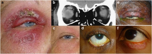
Purpose: Untreated periocular discoid lupus erythematosus (DLE), though very rare, may lead to significant morbidity with lid deformities, trichiasis, and symblepharon formation. We present the largest reported cohort of patients with biopsy-proven DLE solely affecting the periorbital region.
Methods: Observational case series of patients managed over a 7-year period (2004-10).
Results: Seven patients (one male) presented to the Adnexal Service at Moorfields Eye Hospital at a median age of 47 years (range 23-71 years); median interval from symptom onset to biopsy-proven diagnosis was 38 months (range 6-86 months). Changes in peripheral skin were present in 1 patient (occurring after the initial eyelid presentation) and the presenting periocular features were dissimilar across the group, these included: chronic blepharo-conjunctivitis, madarosis, atypical chalazia, depigmentation of the eyelid margin, or marked, persistent periocular oedema with dacryoadenitis.Two cases settled spontaneously, but five required systemic hydroxychloroquine or intralesional corticosteroid injections.
Conclusion: Periorbital DLE is rare and very varied in its presentation, the protean manifestations often resulting in significant diagnostic delay. All patients with unusual periocular skin disease and those with a refractory inflammatory dermopathy, should undergo biopsy of involved tissue(s), thus leading to earlier diagnosis and prevention of permanent cicatricial periocular changes.
Figures
Similar articles
-
Discoid lupus erythematosus masquerading as chronic blepharoconjunctivitis.Ophthalmology. 2005 May;112(5):e19-23. doi: 10.1016/j.ophtha.2005.01.035. Ophthalmology. 2005. PMID: 15878050
-
Chronic blepharitis like picture in patients with Discoid lupus erythematosis - Case series.Nepal J Ophthalmol. 2017 Jul;9(18):175-179. doi: 10.3126/nepjoph.v9i2.19264. Nepal J Ophthalmol. 2017. PMID: 29634708
-
Discoid Lupus Erythematosus Presenting as Upper Eyelid Edema and Erythema.Acta Med Iran. 2017 Jul;55(7):474-476. Acta Med Iran. 2017. PMID: 28918619
-
Unilateral tumid lupus erythematosus.Lupus. 2002;11(6):388-91. doi: 10.1191/0961203302lu208cr. Lupus. 2002. PMID: 12139378 Review.
-
Successful treatment of generalized discoid lupus erythematosus with imiquimod cream 5%: a case report and review of the literature.Acta Dermatovenerol Croat. 2014;22(2):150-9. Acta Dermatovenerol Croat. 2014. PMID: 25102804 Review.
Cited by
-
A Rare Manifestation of Discoid Lupus Erythematosus Solely in the Lower Eyelid of a Young Man.Cureus. 2023 Oct 13;15(10):e47002. doi: 10.7759/cureus.47002. eCollection 2023 Oct. Cureus. 2023. PMID: 37965392 Free PMC article.
-
Periorbital discoid lupus erythematosus: A retrospective study.JAAD Case Rep. 2022 Jun 2;25:78-82. doi: 10.1016/j.jdcr.2022.05.024. eCollection 2022 Jul. JAAD Case Rep. 2022. PMID: 35783074 Free PMC article. No abstract available.
-
Ocular manifestations of systemic lupus erythematosus: a review of the literature.Autoimmune Dis. 2012;2012:290898. doi: 10.1155/2012/290898. Epub 2012 Jul 2. Autoimmune Dis. 2012. PMID: 22811887 Free PMC article.
-
Periocular dermatoses.Int J Womens Dermatol. 2017 Sep 18;3(4):206-218. doi: 10.1016/j.ijwd.2017.08.001. eCollection 2017 Dec. Int J Womens Dermatol. 2017. PMID: 29234715 Free PMC article. Review.
-
Cicatricial conjunctivitis secondary to discoid lupus erythematosus.JAAD Case Rep. 2020 Aug 29;6(12):1323-1326. doi: 10.1016/j.jdcr.2020.08.027. eCollection 2020 Dec. JAAD Case Rep. 2020. PMID: 33299916 Free PMC article. No abstract available.
References
-
- Huey C, Jakobiec FA, Iwamoto T, Kennedy R, Farmer ER, Green WR. Discoid lupus erythematosus of the eyelids. Ophthalmology. 1983;90:1389–1398. - PubMed
-
- Pandhi D, Singal A, Rohtagi J. Eyelid involvement in disseminated chronic cutaneous lupus erythematosus. Indian J Dermatol Venereol Leprol. 2006;72:370–372. - PubMed
-
- Donzis PB, Insler MS, Buntin DM, Gately LE. Discoid lupus erythematosus involving the eyelids. Am J Ophthalmol. 1984;98:32–36. - PubMed
-
- Williams WL, Ramos-Caro FA. Acute periorbital mucinosis in discoid lupus erythematosus. J Am Acad Dermatol. 1999;41:871–873. - PubMed
MeSH terms
Substances
LinkOut - more resources
Full Text Sources
Medical
