. 2012 Mar;14(3):313-22.
doi: 10.1038/gim.2011.13. Epub 2012 Jan 5.

NIPBL rearrangements in Cornelia de Lange syndrome: evidence for replicative mechanism and genotype-phenotype correlation

Affiliations

NIPBL rearrangements in Cornelia de Lange syndrome: evidence for replicative mechanism and genotype-phenotype correlation

Davut Pehlivan et al. Genet Med. 2012 Mar.

Abstract

Purpose: Cornelia de Lange syndrome (CdLS) is a multisystem congenital anomaly disorder characterized by mental retardation, limb abnormalities, distinctive facial features, and hirsutism. Mutations in three genes involved in sister chromatid cohesion, NIPBL, SMC1A, and SMC3, account for ~55% of CdLS cases. The molecular etiology of a significant fraction of CdLS cases remains unknown. We hypothesized that large genomic rearrangements of cohesin complex subunit genes may play a role in the molecular etiology of this disorder.

Methods: Custom high-resolution oligonucleotide array comparative genomic hybridization analyses interrogating candidate cohesin genes and breakpoint junction sequencing of identified genomic variants were performed.

Results: Of the 162 patients with CdLS, for whom mutations in known CdLS genes were previously negative by sequencing, deletions containing NIPBL exons were observed in 7 subjects (~5%). Breakpoint sequences in five patients implicated microhomology-mediated replicative mechanisms-such as serial replication slippage and fork stalling and template switching/microhomology-mediated break-induced replication-as a potential predominant contributor to these copy number variations. Most deletions are predicted to result in haploinsufficiency due to heterozygous loss-of-function mutations; such mutations may result in a more severe CdLS phenotype.

Conclusion: Our findings suggest a potential clinical utility to testing for copy number variations involving NIPBL when clinically diagnosed CdLS cases are mutation-negative by DNA-sequencing studies.


Figures
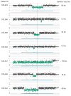
Figure 1
Figure 1. Array comparative genomic hybridization (aCGH) results displaying the NIPBL gene region
Results for aCGH analyses in each of the seven Cornelia de Lange syndrome (CdLS) cases are shown. Individual dots represent interrogating oligonucleotide probes: black dot represents normal copy number as compared with a gender-matched control, red dots represent copy number gain, and green dots represent copy number losses as compared with a gender-matched control. Horizontal blue bar represents the NIPBL gene. Numbers on the y axis show the log2 ratio of the hybridization signal of patient versus control. The approximate size in kb of the deletion is shown at right.
Figure 2
Figure 2. Schematic view of NIPBL displaying exonic deletions in seven patients
(a) Chromosome 5 karyogram with G bands indicated (top). The location of NIPBL is demarcated with a blue vertical line at 5p13.1. (b) Graphic view of 47 exons (vertical black bars) of NIPBL; size and orientation of the gene above the exons. (c) Solid green bars represent genomic regions deleted with approximate sizes. Vertical dotted lines track exons on deleted regions. Patients’ code and deleted exons (Δ) are given at the left. The graphical normalized data for each patient was obtained by inputting the most distal and proximal oligonucleotide genomic probe coordinates into the custom track at the University of California, Santa Cruz website, http://genome.ucsc.edu/cgi-bin/hgGateway. Narrow green vertical bars depict uncertainty for proximal and distal ends of the deleted regions in patients CDL341 and CDL454, for which breakpoint junctions were not determined. Note the 77-kb length of intron 1; thus intron 1 harbors the distal breakpoint in five of seven cases.
Figure 3
Figure 3. Breakpoint sequence analyses for patients with NIPBL deletions
The proximal and distal sequences refer to reference sequences and to their relative position from the centromere. Proximal reference sequence and patient breakpoint sequences that match with the proximal reference sequence are shown in green, whereas the distal reference sequence and patient breakpoint sequences that match with the distal reference sequence are shown in red. Dash boxed sequences (purple) correspond to regions of microhomology and reveal the breakpoint junctions. Patient identification numbers, the type of the repeat sequence, and observed microhomology are shown above.
Figure 4
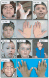
Figure 4. Patient photographs
Frontal view of facies and extremity pictures of patients with limb abnormality.

References

    1. Jackson L, Kline AD, Barr MA, Koch S. de Lange syndrome: a clinical review of 310 individuals. Am J Med Genet. 1993;47:940–946. - PubMed
    1. Kline AD, Barr M, Jackson LG. Growth manifestations in the Brachmann-de Lange syndrome. Am J Med Genet. 1993;47:1042–1049. - PubMed
    1. de Lange C. Sur un type nouveau de degeneration (typus Amstelodamensis) Arch Med Enfants. 1933;36:713–719.
    1. Allanson JE, Hennekam RC, Ireland M. De Lange syndrome: subjective and objective comparison of the classical and mild phenotypes. J Med Genet. 1997;34:645–650. - PMC - PubMed
    1. Krantz ID, McCallum J, DeScipio C, et al. Cornelia de Lange syndrome is caused by mutations in NIPBL, the human homolog of Drosophila melanogaster Nipped-B. Nat Genet. 2004;36:631–635. - PMC - PubMed

Publication types

MeSH terms