Review
. 2012 Jun;33(6):1007-13.
doi: 10.3174/ajnr.A2798. Epub 2012 Jan 12.

Intracranial dural arteriovenous fistulas: classification, imaging findings, and treatment

Affiliations
Review

Intracranial dural arteriovenous fistulas: classification, imaging findings, and treatment

D Gandhi et al. AJNR Am J Neuroradiol. 2012 Jun.

Abstract

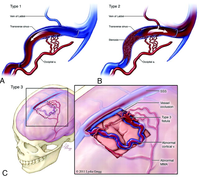
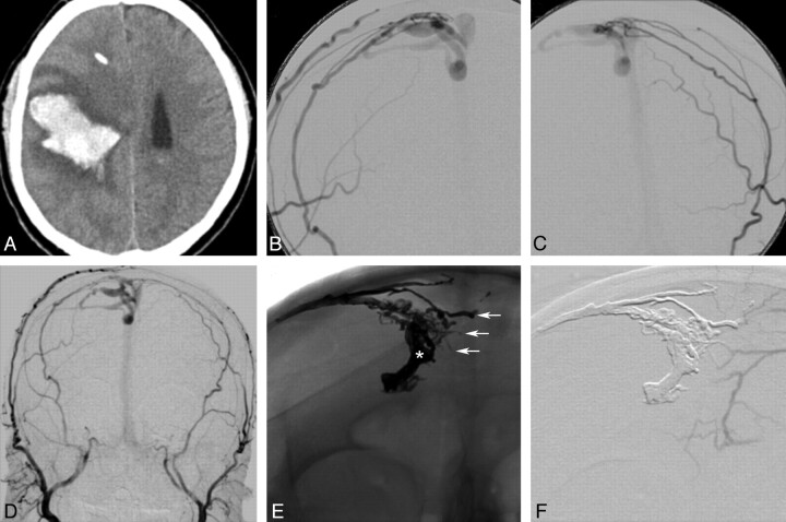
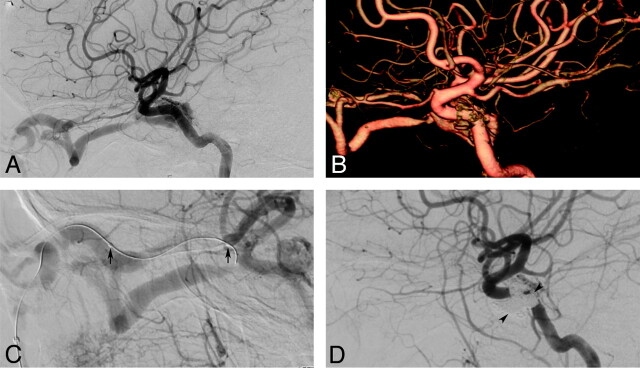
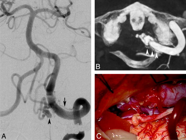
Intracranial DAVFs are pathologic dural-based shunts and account for 10%-15% of all intracranial arteriovenous malformations. These malformations derive their arterial supply primarily from meningeal vessels, and the venous drainage is either via dural venous sinuses or through the cortical veins. DAVFs have a reported association with dural sinus thrombosis, venous hypertension, previous craniotomy, and trauma, though many lesions are idiopathic. The diagnosis is dependent on a high level of clinical suspicion and high-resolution imaging. Cross-sectional imaging techniques by using CT and MR imaging aid in the diagnosis, but conventional angiography remains the most accurate method for complete characterization and classification of DAVFs. The pattern of venous drainage observed on dynamic vascular imaging determines the type of DAVF and correlates with the severity of symptoms and the risk of hemorrhage.


Figures

Fig 1.
Fig 1.
A 53-year-old man with a history of head trauma presented with seizures and right-arm weakness. MR imaging (not shown) demonstrated bilateral parietal hemorrhage and thrombosis of the sagittal sinus. A–C, Sequential sagittal maximum intensity projections from a 320-section dynamic CTA demonstrate a subtle DAVF of the superior sagittal sinus (SSS) supplied by the anterior division of the left middle meningeal artery (arrows). Note the early opacification of the SSS. Venous phase image (C) demonstrates multiple filling defects (asterisk) in the SSS, consistent with SSS thrombosis. D–F, Correlative DSA of the left external carotid artery confirms the DAVF.
Fig 2.
Fig 2.
Schematic overview of the Borden system of classification for DAVFs. A, Type 1 fistula with multiple communications between the occipital artery and transverse sinus. Note antegrade flow and no cortical venous reflux. B, Type 2 lesions are associated with cortical venous reflux. This illustration demonstrates the presence of a transverse sinus fistula with stenosis of the distal transverse and proximal sigmoid sinuses. There is retrograde blood flow into the proximal transverse sinus and the cortical vein (please also see Fig 1E, -F). C, Type 3 fistulas represent communication between the meningeal arteries and cortical vein (or an isolated segment of venous sinus). In this schematic drawing, the cortical vein harboring the fistula near the frontal convexity is tortuous and has multifocal stenoses in its pathway, 2011 Lydia Gregg.
Fig 3.
Fig 3.
A 52-year-old man presented with severe headache, slurred speech, and acute left hemiparesis. A, Noncontrast CT reveals a large right frontoparietal hematoma with intraventricular extension. B–D, A DAVF was suspected on MR imaging (not shown), and DSA was performed. Right (B), left (C), and bilateral (D) external carotid artery injections confirm a convexity DAVF (Borden type 3) with arterial supply from the bilateral middle meningeal and superficial temporal arteries. Transarterial treatment was planned by using the right middle meningeal artery approach (D). A microcatheter was navigated close to the fistula, and the embolization was performed with 2.3 mL of Onyx (E). Note the penetration of Onyx into the arteriovenous junction and the proximal vein (asterisk) as well as arterioarterial reflux (arrows) into the contralateral feeders. F, Ipsilateral (not shown) and contralateral external carotid artery injections confirm complete occlusion of the fistula. The patient made a remarkable recovery during the next 3 months and has mild residual left-arm weakness.
Fig 4.
Fig 4.
A 43-year-old woman with Factor V Leiden mutation developed a spontaneous left-sided dural type (indirect) carotid cavernous fistula (CCF) with proptosis, chemosis, and left cranial nerve VI paralysis. A and B, Lateral view of the left internal carotid artery (ICA) injection and a left ICA 3D reconstruction demonstrates a moderate-flow CCF supplied from multiple left cavernous ICA branches. Additional supply also originated from left external carotid artery (ECA) and right ECA branches (not shown). Given the small size and multiplicity of feeders, transvenous treatment was favored. C and D, A microcatheter was navigated from the left facial vein into the left superior ophthalmic vein (arrows). The cavernous sinus was filled with coils (arrowhead), resulting in complete obliteration of the fistula (D). The proptosis and chemosis resolved the same day, and improvement of left cranial nerve VI function was already evident on postoperative day 1.
Fig 5.
Fig 5.
A 58-year-old male patient presented with thunderclap headache and a small subarachnoid hemorrhage centered at the foramen magnum. A, Left vertebral angiogram demonstrates a dural fistula at the foramen magnum adjacent to the V4 segment of the left vertebral artery. A dominant feeder is noted arising from the left vertebral artery (arrow), and there is a focal venous varix (arrowhead). The venous drainage was along the perimesencephalic vein into the left superior petrosal sinus (not shown). B, Axial DynaCT (Siemens, Erlangen, Germany) reconstruction reveals multiple additional feeders (arrows) that were difficult to appreciate on 2D DSA. The patient underwent an embolization of the larger feeder transarterially, but complete occlusion of the fistula was not accomplished. C, The residual fistula was surgically occluded. A partial C1 laminectomy and suboccipital craniotomy were fashioned. Surgical disconnection of the venous outflow was performed, resulting in complete obliteration of the fistula. The patient made an uncomplicated and complete recovery.

References

    1. Kwon BJ, Han MH, Kang HS, et al. . MR imaging findings of intracranial dural arteriovenous fistulas: relations with venous drainage patterns. AJNR Am J Neuroradiol 2005;26:2500–07 - PMC - PubMed
    1. Kirsch M, Liebig T, Kuhne D, et al. . Endovascular management of dural arteriovenous fistulas of the transverse and sigmoid sinus in 150 patients. Neuroradiology 2009;51:477–83. Epub 2009 Apr 8 - PubMed
    1. Morita A, Meyer FB, Nichols DA, et al. . Childhood dural arteriovenous fistulae of the posterior dural sinuses: three case reports and literature review. Neurosurgery 1995;37:1193–99, discussion 1199–1200 - PubMed
    1. Chung SJ, Kim JS, Kim JC, et al. . Intracranial dural arteriovenous fistulas: analysis of 60 patients. Cerebrovasc Dis 2002;13:79–88 - PubMed
    1. Nabors MW, Azzam CJ, Albanna FJ, et al. . Delayed postoperative dural arteriovenous malformations: report of two cases. J Neurosurg 1987;66:768–72 - PubMed

MeSH terms