. 2012 Sep 1;72(12):1374-81.
doi: 10.1002/pros.22487. Epub 2012 Jan 12.

Effect of dual inhibition of apoptosis and autophagy in prostate cancer

Affiliations

Effect of dual inhibition of apoptosis and autophagy in prostate cancer

Ahamed Saleem et al. Prostate. .

Abstract

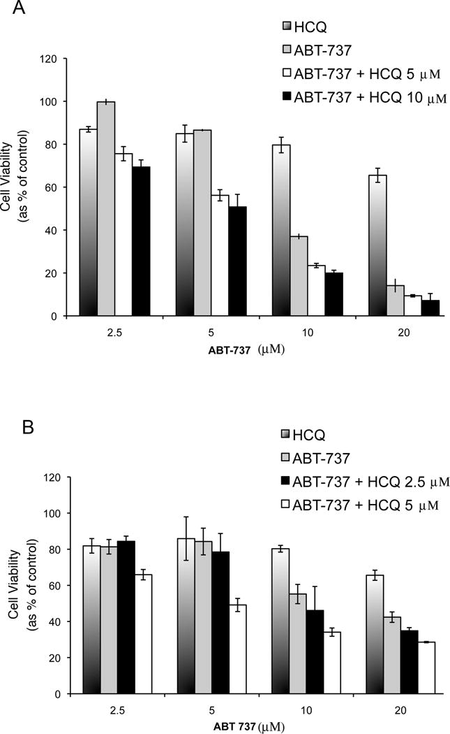
Purpose: Targeting multiple anti-apoptotic proteins is now possible with the small molecule BH3 domain mimetics such as ABT-737. Given recent studies demonstrating that autophagy is a resistance mechanism to multiple therapeutic agents in the setting of apoptotic inhibition, we hypothesized that hydroxychloroquine (HCQ), an anti-malarial drug that inhibits autophagy, will increase cytotoxicity of ABT-737.

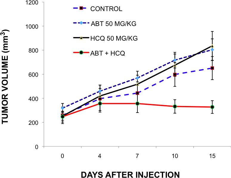
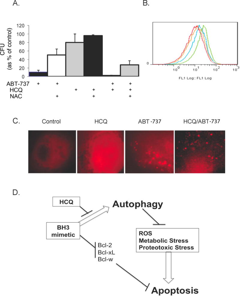
Experimental design: Cytotoxicity of ABT-737 and HCQ was assessed in vitro in PC-3 and LNCaP cells, and in vivo in a xenograft mouse model. The role of autophagy as a resistance mechanism was assessed by siRNA knockdown of the essential autophagy gene beclin1. ROS was measured by flow cytometry, and mitophagy assessed by the mCherry-Parkin reporter.

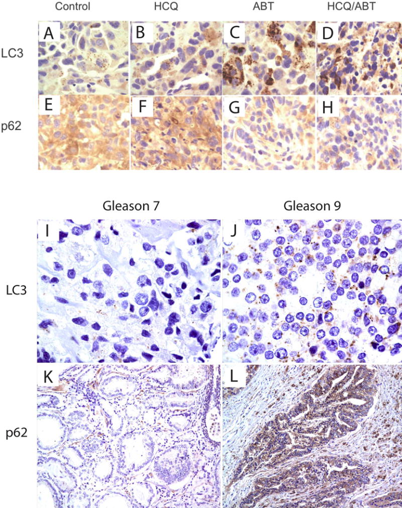
Results: Induction of autophagy by ABT-737 was a mechanism of resistance in prostate cancer cell lines. Therapeutic inhibition of autophagy with HCQ increased cytotoxicity of ABT-737 both in vitro and in vivo. ABT-737 induced LC-3 and decreased p62 expression by immunoblot in cell lines and by immunohistochemistry in tumors in vivo. Assessment of ROS and mitochondria demonstrated that ROS production by ABT-737 and HCQ was a mechanism of cytotoxicity.

Conclusions: We demonstrated that autophagy inhibition with HCQ enhances ABT-737 cytotoxicity in vitro and in vivo, that LC-3 and p62 represent assessable markers in human tissue for future clinical trials, and that ROS induction is a mechanism of cytotoxicity. These results support a new paradigm of dual targeting of apoptosis and autophagy in future clinical studies.


Figures

Figure 1
Figure 1
Cell viability assay with and without inhibition of autophagy. 1A: PC-3 cells transfected with EGFP-LC3 reporter treated with 10uM of Hydroxychloroquine (HCQ), or 5uM of ABT-737 for 72 hours demonstrating LC-3 localized to autophagosomes by fluorescence microscopy. 1B: Immunoblot of LC3-I, LC3-II, and p62 expression in LNCaP and PC3 cells treated with HCQ or ABT-737. The ratio of LC3-II/I represents an assessment of cleavage of LC3 with autophagosome formation. 1C: PC-3 and LNCaP cells were treated for 72 hours with 2.5uM of ABT-737 in cells transfected with beclin1 siRNA or Lamin siRNA control. The X axis of Figure 1C represents treatment of cells with ABT-737 with either Beclin siRNA to decrease Beclin1 (shown as negative (−) for Beclin1) or with Lamin siRNA (shown as positive (+) for Beclin1) in PC-3 and LNCaP cells respectively. The effect of drug treatment on cell viability was determined by MTT assay. The percentage of cell viability as a percentage of untreated cells is shown to decrease with treatment of ABT-737 in both PC-3 and LNCaP cells, with a greater decrease in cell viability with Beclin1 inhibition. All experiments were conducted in triplicate.
Figure 2
Figure 2
Cell viability assay of PC-3 (A) and LNCaP cells (B) in culture treated with HCQ alone, ABT-263 alone at indicated concentrations, and the combination at differing ABT-263 concentrations. Cells were seeded in 96-well plates 24 hours before treatment. MTT assays were performed 48 hours after treatment. The percentage of cell viability is shown for each treatment group compared to the viability of untreated cells, which were considered as 100%. Experiments were conducted in triplicate.
Figure 3
Figure 3
Effect of ABT-737 (ABT), Hydroxychloroquine (HCQ), and the combination in vivo. In vivo tumor growth is shown with combined ABT-737 and HCQ treatment in mouse xenograft model. PC-3 prostate cancer cells were injected subcutaneously in nude mice and grown for 10 days. Mice were treated daily for 14 days with vehicle control, ABT-737 (50mgm/kg), HCQ (50mgm/kg) or both in combination. Each treatment was performed in quadruplicate.
Figure 4
Figure 4
Assessment of LC-3 and p62 in human tumor xenograft and human tissue. 4A-H: Histologic sections were obtained from the tumors of the mice in each treatment group from xenograft studies. LC3 and p62 antibody staining was performed in the tumor tissue sections from Control (A and E), ABT-737 (C and G), HCQ (B and F) and combination of ABT-737 and HCQ (D and H) by immunohistochemistry at 14 days of treatment. 4I–L: Assessment of LC3 and p62 in human cancers with low (Gleason Score 7) and high (Gleason Score 9) grade tumors.
Figure 5
Figure 5
Effect of ABT-737 and HCQ on ROS and mitochondrial function. 5A: PC-3 cells were treated with 10uM of ABT-737 alone and in combination with 1mM N-acetylcysteine (first and second bars on the left) and assessed by clonogenic assay; treated with HCQ alone and in combination with N-acetylcysteine (middle two bars); or treated with the combination of both ABT-737 and HCQ with and without N-acetylcysteine (last two bars on right of figure 5A). 5B: ROS levels were determined using 10uM 2′-7′-dichlorodihydrofluorescene diacetate (DCF-DA, Molecular Probes) in cells treated with the combination of ABT-737 and HCQ (green), ABT-737 (blue), HCQ (yellow), or untreated control (red). 5C: mCherry-Parkin expressing PC-3 cells treated with 20uM of HCQ, 10uM of ABT-737 or the combination of ABT-737 and HCQ to identify dysfunctional mitochondria. 5D: a depiction of autophagy as a cell survival and resistance mechanism through abrogation of oxidative (ROS), metabolic and proteotoxic stress. Figure 5D shows a potential paradigm of optimal tumor targeting by dual inhibition of apoptosis and autophagy.

References

    1. Castilla C, Congregado B, Chinchon D, Torrubia FJ, Japon MA, Saez C. Bcl-xL is overexpressed in hormone-resistant prostate cancer and promotes survival of LNCaP cells via interaction with proapoptotic Bak. Endocrinology. 2006;147:4960–7. - PubMed
    1. Yoshino T, Shiina H, Urakami S, et al. Bcl-2 expression as a predictive marker of hormone-refractory prostate cancer treated with taxane-based chemotherapy. Clin Cancer Res. 2006;12:6116–24. - PubMed
    1. Lin Y, Fukuchi J, Hiipakka RA, Kokontis JM, Xiang J. Up-regulation of Bcl-2 is required for the progression of prostate cancer cells from an androgen-dependent to an androgen-independent growth stage. Cell Res. 2007;17:531–6. - PubMed
    1. Cragg MS, Harris C, Strasser A, Scott CL. Unleashing the power of inhibitors of oncogenic kinases through BH3 mimetics. Nat Rev Cancer. 2009;9:321–6. - PubMed
    1. Chonghaile TN, Letai A. Mimicking the BH3 domain to kill cancer cells. Oncogene. 2008;27(Suppl 1):S149–57. - PMC - PubMed

MeSH terms